ANXA1 Antibodies
Background
The ANXA1 gene encodes the membrane-associated protein A1. This protein belongs to the calcium-dependent phospholipid-binding protein family and is mainly distributed in the cytoplasm and cell membranes of vertebrates. By binding to phospholipids in the cell membrane, this protein participates in regulating key biological processes such as inflammatory responses, cell apoptosis, and membrane repair. In the immune system, ANXA1 acts as an intermediary molecule for the effects of glucocorticoids, inhibiting the migration of white blood cells and exerting anti-inflammatory effects. This gene was first identified in 1986, and the three-dimensional structure analysis further revealed the spatial conformation characteristics of the calcium ion and phospholipid binding domains. The molecular mechanism study of ANXA1 provides an important theoretical basis for understanding immune regulation, tumor occurrence, and tissue repair, and has broad potential in the development of disease treatment targets.
Structure of ANXA1
ANXA1 is a calcium-dependent phospholipid-binding protein with a molecular weight of approximately 37-39 kDa. Its molecular weight varies among different species due to transcript splicing and post-translational modifications.
| Species | Human | Mouse | Rat |
| Molecular Weight (kDa) | ~37 | ~38 | ~39 |
| Primary Structural Differences | Has an N-terminal domain that can be cleaved by proteolysis | Highly conservative, functionally similar | The structures are highly similar, and they are used for research on inflammatory models. |
This protein is composed of approximately 346 amino acids. Its core structure is a conserved C-terminal annexin repeat domain, consisting of four identical α-helical rings, which is responsible for the binding of calcium ions and membrane phospholipids. The N-terminal domain has unique conformational flexibility and contains the glucocorticoid-mediated anti-inflammatory action target and protein interaction interface. The protein binds to the cell membrane through its core domain upon calcium ion triggering and plays a key role in membrane reorganization, signal transduction, and inflammation resolution.
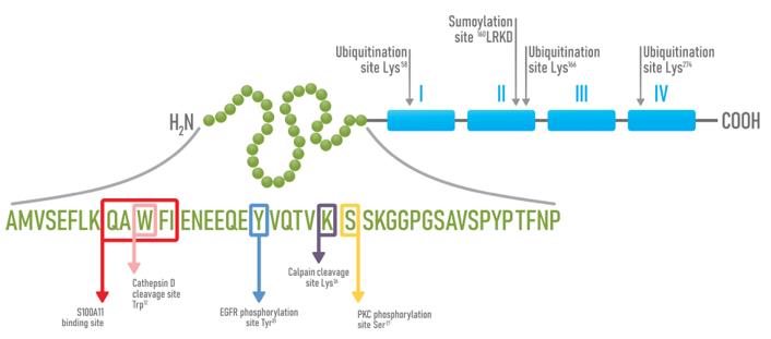 Fig. 1 Schematic representation of AnxA1 structure and its sites of post-translational modifications.1
Key structural properties of ANXA1:
- The core is a flat disc-shaped calcium ion-binding domain, mainly composed of α -helices
- Calcium combined with ring and hydrophobic channel mediates the interaction with the cell membrane
- Variable structure of n-terminal domain with regulation of protein phosphorylation sites
Functions of ANXA1
The main function of ANXA1 is to regulate the inflammatory response and membrane repair processes within cells. At the same time, it is also involved in various physiological and pathological processes such as cell signal transduction, apoptosis regulation, and tissue regeneration.
| Function | Description |
| Inflammation Regulation | As a downstream mediator of glucocorticoids, it inhibits the migration and adhesion of white blood cells and promotes the resolution of inflammation. |
| Membrane Repair | It accumulates at the site of membrane damage in a calcium ion-dependent manner, promoting membrane remodeling and repair. |
| Cell Apoptosis Regulation | Participates in regulating the process of programmed cell death and influences the fate determination of immune cells. |
| Signal Transduction | Interacts with various membrane receptors and signaling molecules, mediating intracellular signaling pathways. |
| Tissue Repair | Promote tissue remodeling and regeneration in the later stage of inflammation, and restore tissue homeostasis. |
The function of ANXA1 is highly dependent on the environment, and its activity shows dynamic changes during the early stage of inflammation, the recovery period, and the repair phase. This characteristic makes it a key molecule that connects the immune response with tissue repair.
Applications of ANXA1 and ANXA1 Antibody in Literature
1. Araújo, Thaise Gonçalves, et al. "Annexin A1 as a regulator of immune response in cancer." Cells 10.9 (2021): 2245. https://doi.org/10.3390/cells10092245
The article indicates that annexin A1 in tumors shifts from anti-inflammatory to pro-inflammatory. It promotes tumor invasion and metastasis through autocrine signaling and by inducing M2-type macrophages, and inhibits anti-tumor immune responses, thereby creating an immunosuppressive microenvironment conducive to tumor growth.
2. Sheikh, Madeeha H., and Egle Solito. "Annexin A1: uncovering the many talents of an old protein." International journal of molecular sciences 19.4 (2018): 1045. https://doi.org/10.3390/ijms19041045
The article indicates that Annexin A1 is not only a crucial anti-inflammatory protein, but also regulates immune repair, endocrine balance and the blood-brain barrier, and affects various pathological processes such as cancer and neurodegenerative diseases, demonstrating potential for diagnosis and treatment.
3. Gong, Yanyu, et al. "ANXA1 promotes intrahepatic cholangiocarcinoma proliferation and growth by regulating glutamine metabolism through GOT1 stabilization." Journal of Experimental & Clinical Cancer Research 44.1 (2025): 151. https://doi.org/10.1186/s13046-025-03400-z
The study found that ANXA1 is highly expressed in intrahepatic cholangiocarcinoma and predicts a poor prognosis. It stabilizes the metabolic enzyme GOT1 by recruiting the deubiquitinating enzyme USP5, promotes glutamine metabolism, reduces oxidative stress, and thereby drives tumor proliferation. Inhibiting ANXA1 and reducing glutamine uptake is a potential therapeutic strategy.
4. Shao, Gang, et al. "Identification of ANXA1 as a Novel Upstream Negative Regulator of Notch1 Function in AML." Advanced Science 11.48 (2024): 2409726. https://doi.org/10.1002/advs.202409726
The study found that in acute myeloid leukemia, the high expression of annexin A1 (ANXA1) inhibits the tumor suppressive function of the Notch1-p15 signaling axis by directly binding to Notch1 protein and promoting its degradation, thereby driving the proliferation of cancer cells. Targeting ANXA1 becomes a potential therapeutic strategy.
5. Li, Wei, et al. "YTHDC1 is downregulated by the YY1/HDAC2 complex and controls the sensitivity of ccRCC to sunitinib by targeting the ANXA1-MAPK pathway." Journal of Experimental & Clinical Cancer Research 41.1 (2022): 250. https://doi.org/10.1186/s13046-022-02460-9
The study found that YTHDC1 is expressed at a low level in renal clear cell carcinoma and predicts a poor prognosis. It slows down tumor progression by inhibiting the ANXA1/MAPK pathway, and regulates the sensitivity of cancer cells to tyrosine kinase inhibitors through the YY1/HDAC2/YTHDC1/ANXA1 signaling axis.
Creative Biolabs: ANXA1 Antibodies for Research
Creative Biolabs specializes in the production of high-quality ANXA1 antibodies for research and industrial applications. Our portfolio includes monoclonal and polyclonal antibodies tailored for ELISA, Flow Cytometry, Western blot, immunohistochemistry, and other diagnostic methodologies.
- Custom ANXA1 Antibody Development: Tailor-made solutions to meet specific research requirements.
- Bulk Production: Large-scale antibody manufacturing for industry partners.
- Technical Support: Expert consultation for protocol optimization and troubleshooting.
- Aliquoting Services: Conveniently sized aliquots for long-term storage and consistent experimental outcomes.
For more details on our ANXA1 antibodies, custom preparations, or technical support, contact us at email.
Reference
- Araújo, Thaise Gonçalves, et al. "Annexin A1 as a regulator of immune response in cancer." Cells 10.9 (2021): 2245. Distributed under Open Access license CC BY 4.0, without modification. https://doi.org/10.3390/cells10092245
Anti-ANXA1 antibodies
 Loading...


Hot products 
-
Mouse Anti-FTH1 Recombinant Antibody (CBXF-1896) (CBMAB-F3426-CQ)

-
Mouse Anti-8-oxoguanine Recombinant Antibody (V2-7719) (CBMAB-1898CQ)

-
Mouse Anti-GFP Recombinant Antibody (28) (CBMAB-G3038-LY)

-
Mouse Anti-A2M Recombinant Antibody (V2-178822) (CBMAB-A0036-YC)

-
Mouse Anti-C5b-9 Recombinant Antibody (aE11) (CBMAB-AO138LY)

-
Mouse Anti-FOXL1 Recombinant Antibody (CBXF-0845) (CBMAB-F0462-CQ)

-
Mouse Anti-CORO1A Recombinant Antibody (4G10) (V2LY-1206-LY806)

-
Mouse Anti-CFL1 (Phospho-Ser3) Recombinant Antibody (CBFYC-1770) (CBMAB-C1832-FY)

-
Mouse Anti-ADGRE2 Recombinant Antibody (V2-261270) (CBMAB-C0813-LY)

-
Mouse Anti-CCT6A/B Recombinant Antibody (CBXC-0168) (CBMAB-C5570-CQ)

-
Mouse Anti-CCNH Recombinant Antibody (CBFYC-1054) (CBMAB-C1111-FY)

-
Mouse Anti-DMPK Recombinant Antibody (CBYCD-324) (CBMAB-D1200-YC)

-
Mouse Anti-FOXA3 Recombinant Antibody (2A9) (CBMAB-0377-YC)

-
Mouse Anti-AAV9 Recombinant Antibody (V2-634029) (CBMAB-AP023LY)

-
Mouse Anti-CSPG4 Recombinant Antibody (CBFYM-1050) (CBMAB-M1203-FY)

-
Mouse Anti-BBS2 Recombinant Antibody (CBYY-0253) (CBMAB-0254-YY)

-
Mouse Anti-AKR1B1 Antibody (V2-2449) (CBMAB-1001CQ)

-
Rabbit Anti-ATF4 Recombinant Antibody (D4B8) (CBMAB-A3872-YC)

-
Rat Anti-CD63 Recombinant Antibody (7G4.2E8) (CBMAB-C8725-LY)

-
Rat Anti-FABP3 Recombinant Antibody (CBXF-2299) (CBMAB-F1612-CQ)

- AActivation
- AGAgonist
- APApoptosis
- BBlocking
- BABioassay
- BIBioimaging
- CImmunohistochemistry-Frozen Sections
- CIChromatin Immunoprecipitation
- CTCytotoxicity
- CSCostimulation
- DDepletion
- DBDot Blot
- EELISA
- ECELISA(Cap)
- EDELISA(Det)
- ESELISpot
- EMElectron Microscopy
- FFlow Cytometry
- FNFunction Assay
- GSGel Supershift
- IInhibition
- IAEnzyme Immunoassay
- ICImmunocytochemistry
- IDImmunodiffusion
- IEImmunoelectrophoresis
- IFImmunofluorescence
- IGImmunochromatography
- IHImmunohistochemistry
- IMImmunomicroscopy
- IOImmunoassay
- IPImmunoprecipitation
- ISIntracellular Staining for Flow Cytometry
- LALuminex Assay
- LFLateral Flow Immunoassay
- MMicroarray
- MCMass Cytometry/CyTOF
- MDMeDIP
- MSElectrophoretic Mobility Shift Assay
- NNeutralization
- PImmunohistologyp-Paraffin Sections
- PAPeptide Array
- PEPeptide ELISA
- PLProximity Ligation Assay
- RRadioimmunoassay
- SStimulation
- SESandwich ELISA
- SHIn situ hybridization
- TCTissue Culture
- WBWestern Blot
