CTNS Antibodies
Background
CTNS gene encoding a membrane protein called cystine transporters, are mainly distributed in the lysosome membrane, is responsible for cystine to dissolve enzyme in vitro to maintain cystine steady state in the cell. The defect of this gene can lead to abnormal accumulation of cystine in lysosomes, causing a rare cystine storage disorder, mainly manifested as cystine crystal deposition in multiple organs and progressive tissue damage. In 1998, multiple research teams collaborated to complete the localization cloning, and the study of its pathogenic mechanism provided an important model for lysosomal storage disorders. As the first lysosomal membrane protein target to conduct clinical trials using gene therapy, the study of the molecular characteristics and functions of the CTNS gene not only deepens people's understanding of amino acid transport mechanisms but also lays the foundation for the development of treatment strategies for various genetic metabolic diseases.
Structure of CTNS
The cystinosin encoded by the CTNS gene is a lysosomal membrane protein with a molecular weight of approximately 55 kDa, and its size varies slightly among different species:
| Species | Human | Mouse | Rat | Bovine |
| Molecular Weight (kDa) | 55 | 54.8 | 55.2 | 54.9 |
| Primary Structural Differences | Seven transmembrane domains, N-terminal lysosomal targeting signals | Highly conserved, similar to human proteins | Across the membrane area highly homologous | Functional domains are highly conserved |
This protein is composed of 367 amino acids and has 7 transmembrane domains (TM1-TM7). Its N-terminal contains lysosomal targeting signals (GYDQL), ensuring its correct localization on the lysosomal membrane. The key functional sites include the "YFPQA" motif located at TM7, which is responsible for cystine transport, while the C-terminal tail segment (residues 318-367) within the lysosomal lumen is involved in regulating protein stability. Mutations in this protein can lead to cystine storage disorder, characterized by the accumulation of cystine crystals in lysosomes, which affects the functions of multiple organs such as the kidneys and eyes.
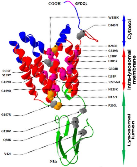 Fig. 1 Molecular mechanism of CTNS gene mutation and functional defect of cystine transporter.1
Key structural properties of CTNS:
- 7-transmembrane helical structure (TM1-TM7)
- N-terminal lysosomal targeting signal (GYDQL sequence)
- Conserved "YFPQA" motif (located in TM7)
- C-terminal lysosomal lumen domain (Residues 318-367)
- Mutation hotspot regions (such as exon 3-5)
Functions of CTNS
The core function of the protein encoded by the CTNS gene is to maintain cystine homeostasis in lysosomes, and its functional defect is closely related to the pathogenesis of cystine storage disorders.
| Function | Description |
| Cystine transport | Mediate the excretion of cystine within lysosomes and prevent the accumulation of cystine crystals. |
| Lysosomal function is maintained | Ensure the normal metabolism of lysosomes and avoid lysosomal dysfunction caused by cystine deposition. |
| Cellular REDOX balance | By adjusting the level of cystine indirectly affect GSH synthesis, involved in antioxidant defense. |
| Multi-organ protection | Prevent tissue damage caused by cystine crystallization in organs such as the kidneys, corneas, and thyroid. |
| Autophagy regulation | Lysosomal cystine accumulation may interfere with autophagic flux and affect cellular waste clearance. |
This protein relies on the lysosomal membrane potential and pH gradient for the active transport of cystine. The loss of its function will cause the cystine concentration in lysosomes to increase by more than 100 times, resulting in the formation of characteristic crystals. Unlike the synergistic effect of hemoglobin, the function of CTNS protein is cell-specific, especially playing a crucial role in proximal renal tubular epithelial cells, and its mutation can trigger Fanconi syndrome.
Applications of CTNS and CTNS Antibody in Literature
1. Elmonem, Mohamed A., et al. "Cystinosis: a review." Orphanet journal of rare diseases 11.1 (2016): 47.https://doi.org/10.1186/s13023-016-0426-y
This article reviews cystine storage syndrome caused by CTNS gene mutations, which leads to the accumulation of cystine in lysosomes and causes multiple organ damage. The clinical manifestations, diagnostic methods (such as cystine detection in white blood cells) and treatment strategies (such as cysteine) were mainly discussed, and the future research directions were prospected.
2. Elmonem, Mohamed A., et al. "Cystinosis (ctns) zebrafish mutant shows pronephric glomerular and tubular dysfunction." Scientific reports 7.1 (2017): 42583.https://doi.org/10.1038/srep42583
This article indicates that mutations in the CTNS gene lead to functional defects of cystine transporters, causing lysosomal cystine accumulation and cystine storage disorders. Researchers simulated human kidney lesions through zebrafish models (ctns mutants) and confirmed that cysteamine can alleviate apoptosis, providing a new tool for mechanism research and treatment development.
3. Chkioua, Latifa, et al. "Molecular characterization of CTNS mutations in Tunisian patients with ocular cystinosis." Diagnostic Pathology 17.1 (2022): 44.https://doi.org/10.1186/s13000-022-01221-8
Research through corneal OCT and gene sequencing has found that CTNS gene mutations (such as p.S139Y, p.Q88K, etc.) lead to abnormal function of cystine transporters, causing cystine crystal deposition in the eyes. Novel transmembrane region mutations may cause disease by affecting protein stability, and cysteamine treatment can improve symptoms.
4. Reda, Ahmed, Koenraad Veys, and Martine Besouw. "Fertility in cystinosis." Cells 10.12 (2021): 3539.https://doi.org/10.3390/cells10123539
This article reviews the fertility issues of patients with cystine storage disorder (CTNS gene defect), focusing on the differences in infertility caused by azoospermia in male patients and the fertility of female patients. It analyzes the impact of cysteamine treatment on fertility function and points out the deficiencies of current research and future directions.
5. Papizh, Svetlana, et al. "CTNS mRNA molecular analysis revealed a novel mutation in a child with infantile nephropathic cystinosis: a case report." BMC nephrology 20.1 (2019): 400.https://doi.org/10.1186/s12882-019-1589-2
This study reports a case of infantile renal cysteine storage syndrome born from consanguineous marriage. RT-PCR and cDNA sequencing revealed a novel 9kb homozygous deletion (spanning introns 3 to 5) in the CTNS gene, and both parents were heterozygous carriers. RNA level analysis is helpful in clarifying the genetic defects of such patients.
Creative Biolabs: CTNS Antibodies for Research
Creative Biolabs specializes in the production of high-quality CTNS antibodies for research and industrial applications. Our portfolio includes monoclonal antibodies tailored for ELISA, Flow Cytometry, Western blot, immunohistochemistry, and other diagnostic methodologies.
- Custom CTNS Antibody Development: Tailor-made solutions to meet specific research requirements.
- Bulk Production: Large-scale antibody manufacturing for industry partners.
- Technical Support: Expert consultation for protocol optimization and troubleshooting.
- Aliquoting Services: Conveniently sized aliquots for long-term storage and consistent experimental outcomes.
For more details on our CTNS antibodies, custom preparations, or technical support, contact us at email.
Reference
- Chkioua, Latifa, et al. "Molecular characterization of CTNS mutations in Tunisian patients with ocular cystinosis." Diagnostic Pathology 17.1 (2022): 44.https://doi.org/10.1186/s13000-022-01221-8
Anti-CTNS antibodies
 Loading...


Hot products 
-
Mouse Anti-ABCA3 Recombinant Antibody (V2-178911) (CBMAB-A0145-YC)

-
Rat Anti-CD63 Recombinant Antibody (7G4.2E8) (CBMAB-C8725-LY)

-
Mouse Anti-FYN Recombinant Antibody (10) (CBMAB-S6332-CQ)

-
Mouse Anti-CD33 Recombinant Antibody (6C5/2) (CBMAB-C8126-LY)

-
Mouse Anti-AAV-5 Recombinant Antibody (V2-503416) (CBMAB-V208-1402-FY)

-
Mouse Anti-C1QC Recombinant Antibody (CBFYC-0600) (CBMAB-C0654-FY)

-
Rat Anti-ADGRE4 Recombinant Antibody (V2-160163) (CBMAB-F0011-CQ)

-
Mouse Anti-CECR2 Recombinant Antibody (CBWJC-2465) (CBMAB-C3533WJ)

-
Mouse Anti-FPR2 Recombinant Antibody (1D6) (CBMAB-F2628-CQ)

-
Mouse Anti-CAPZB Recombinant Antibody (CBYY-C0944) (CBMAB-C2381-YY)

-
Mouse Anti-CD19 Recombinant Antibody (CBXC-1224) (CBMAB-C1491-CQ)

-
Mouse Anti-ARID3A Antibody (A4) (CBMAB-0128-YC)

-
Mouse Anti-AAV9 Recombinant Antibody (V2-634029) (CBMAB-AP023LY)

-
Mouse Anti-GFAP Recombinant Antibody (5) (CBMAB-G0346-LY)

-
Mouse Anti-APCS Recombinant Antibody (CBYC-A663) (CBMAB-A3054-YC)

-
Rabbit Anti-CCN1 Recombinant Antibody (CBWJC-3580) (CBMAB-C4816WJ)

-
Mouse Anti-CD46 Recombinant Antibody (CBFYC-0076) (CBMAB-C0085-FY)

-
Mouse Anti-DLG1 Monolconal Antibody (4F3) (CBMAB-0225-CN)

-
Mouse Anti-DHFR Recombinant Antibody (D0821) (CBMAB-D0821-YC)

-
Mouse Anti-CD83 Recombinant Antibody (HB15) (CBMAB-C1765-CQ)

- AActivation
- AGAgonist
- APApoptosis
- BBlocking
- BABioassay
- BIBioimaging
- CImmunohistochemistry-Frozen Sections
- CIChromatin Immunoprecipitation
- CTCytotoxicity
- CSCostimulation
- DDepletion
- DBDot Blot
- EELISA
- ECELISA(Cap)
- EDELISA(Det)
- ESELISpot
- EMElectron Microscopy
- FFlow Cytometry
- FNFunction Assay
- GSGel Supershift
- IInhibition
- IAEnzyme Immunoassay
- ICImmunocytochemistry
- IDImmunodiffusion
- IEImmunoelectrophoresis
- IFImmunofluorescence
- IGImmunochromatography
- IHImmunohistochemistry
- IMImmunomicroscopy
- IOImmunoassay
- IPImmunoprecipitation
- ISIntracellular Staining for Flow Cytometry
- LALuminex Assay
- LFLateral Flow Immunoassay
- MMicroarray
- MCMass Cytometry/CyTOF
- MDMeDIP
- MSElectrophoretic Mobility Shift Assay
- NNeutralization
- PImmunohistologyp-Paraffin Sections
- PAPeptide Array
- PEPeptide ELISA
- PLProximity Ligation Assay
- RRadioimmunoassay
- SStimulation
- SESandwich ELISA
- SHIn situ hybridization
- TCTissue Culture
- WBWestern Blot
