
CXCL8 Antibodies
Background
CXCL8 is a small molecule chemokine protein mainly secreted by immune cells and belongs to the CXC chemokine family. The protein encoded by this gene can specifically bind to and activate IL-8 receptors (such as CXCR1/CXCR2) on the cell surface, inducing the directional migration and activation of immune cells such as neutrophils, thereby playing a core role in inflammatory responses and immune defense. This gene was first identified by multiple research teams in 1986 and is the first human chemokine confirmed to have neutrophil chemotactic function. Its gene expression is precisely regulated by inflammatory pathways such as NF-κB, and the mechanism of its three-dimensional structure interacting with receptors has been deeply analyzed, providing an important target research basis for the development of anti-inflammatory drugs.
Structure of CXCL8
CXCL8 (interleukin-8) is a small chemokine protein with a molecular weight of approximately 8.4 kDa. There are slight differences in its molecular weight among different species, mainly due to changes in amino acid sequences.
| Species | Human | Mouse | Rat |
| Molecular Weight (kDa) | 8.4 | 8.0 | 8.2 |
| Primary Structural Differences | Contains 72 amino acids, core ELR motif | Lack of ELR motif, functional differentiation | Sequence highly homologous with humans |
This protein is composed of 72 amino acids, and its three-dimensional structure presents a typical chemokine folding, consisting of three antiparallel β -sheets and a C-terminal α -helix. The N-terminal of the protein contains a key "glutamate-leucine-arginine" (ELR) motif, which is the core functional domain through which it binds to the receptor CXCR1/CXCR2 and mediates the chemotactic activity of neutrophils. The positively charged residues on its surface help interact with glycosaminoglycans (GAGs) on the cell surface, thereby forming a local chemical concentration gradient.
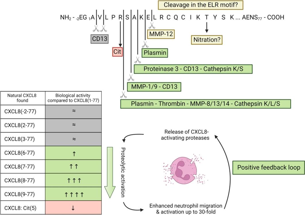 Fig. 1 Post-translational modifications affect the biological activity of CXCL8.1
Key structural properties of CXCL8:
- Stable three-strand anti-parallel β -folded structure
- N-terminal "ELR" motifs mediate receptor specific recognition and activation
- Amino acid residues with positive charges on the surface combine with glycosaminoglycans
- C-terminal α-helical protein conformation stabilization and influence chemotactic function
Functions of CXCL8
The primary function of CXCL8 (interleukin-8) is to chemotaxis and activate neutrophils, but it is also widely involved in pathophysiological processes such as inflammatory responses, angiogenesis, and tumorigenesis.
| Function | Description |
| Neutrophil chemotaxis | As a potent chemokine, it binds to receptors CXCR1/CXCR2 and guides neutrophils to migrate to the inflammatory site. |
| Angiogenesis regulation | In tumor microenvironment promote new blood vessel formation, various conditions such as support for tissue repair and tumor growth. |
| Amplification of inflammatory signals | Stimulate neutrophil degranulation, release reactive oxygen species and enzymes, and enhance local inflammatory responses. |
| Activation of immune cells | In addition to neutrophils, it can also chemotactic and partially activate other immune cells such as T cells and monocytes. |
| Pathogen defense | By recruiting phagocytes to the infected site, the body's ability to clear bacteria and other pathogens is enhanced. |
CXCL8 exerts its function through the G protein-coupled receptor signaling pathway, and its expression is strictly regulated by transcription factors such as NF-κB and AP-1. It often shows persistently high expression in chronic inflammation and cancer.
Applications of CXCL8 and CXCL8 Antibody in Literature
1. Cambier, Seppe, Mieke Gouwy, and Paul Proost. "The chemokines CXCL8 and CXCL12: molecular and functional properties, role in disease and efforts towards pharmacological intervention." Cellular & molecular immunology 20.3 (2023): 217-251. https://doi.org/10.1038/s41423-023-00974-6
The article indicates that CXCL8 is a key chemokine that strongly recruits neutrophils by binding to CXCR1/CXCR2 receptors and glycosaminoglycan, playing a significant role in infections, inflammation and cancer, and is an important target in current therapeutic research.
2. Yang, Qingran, et al. "The CXCL8/MAPK/hnRNP-K axis enables susceptibility to infection by EV-D68, rhinovirus, and influenza virus in vitro." Nature Communications 16.1 (2025): 1715. https://doi.org/10.1038/s41467-025-57094-0
Studies have found that the respiratory virus EV-D68 activates the MAPK pathway by binding CXCL8 to its receptor CXCR1/2, promoting the intracellular entry of hnRNP-K protein in the nucleus and enhancing viral RNA recognition, thereby facilitating viral replication. This mechanism also exists in influenza and rhinoviruses.
3. Gao, Jian, et al. "SKAP1 expression in cancer cells enhances colon tumor growth and impairs cytotoxic immunity by promoting neutrophil extracellular trap formation via the NFATc1/CXCL8 axis." Advanced Science 11.41 (2024): 2403430. https://doi.org/10.1002/advs.202403430
Research has found that SKAP1 upregulates the expression of CXCL8 in colon cancer cells by activating NFATc1, thereby promoting neutrophil infiltration and NET formation, and accelerating tumor progression. Targeting this axis can enhance the anti-tumor effect of NK cell therapy.
4. Bie, Yaqin, et al. "[Retracted] The Crucial Role of CXCL8 and Its Receptors in Colorectal Liver Metastasis." Disease markers 2019.1 (2019): 8023460. https://doi.org/10.1155/2019/8023460
Research has found that CXCL8 promotes the progression, metastasis and immune escape of colorectal cancer by binding to receptors such as CXCR1/CXCR2, and induces epithelial-mesenchymal transition and resistance to nest-loss apoptosis. Targeting this signaling axis is a potential strategy for overcoming drug resistance and inhibiting tumor development.
5. Zhang, et al. "EBV-induced CXCL8 upregulation promotes vasculogenic mimicry in gastric carcinoma via NF-κB signaling." Frontiers in cellular and infection microbiology 12 (2022): 780416. https://doi.org/10.3389/fcimb.2022.780416
Research has found that EBV promotes the formation of angiogenic mimicry in gastric cancer cells by up-regulating the expression of CXCL8 and activating the NF-κB signaling pathway, thereby enhancing their proliferation and migration capabilities. CXCL8 is highly expressed in EBV-related gastric cancer and is a potential therapeutic target.
Creative Biolabs: CXCL8 Antibodies for Research
Creative Biolabs specializes in the production of high-quality CXCL8 antibodies for research and industrial applications. Our portfolio includes monoclonal antibodies tailored for ELISA, Flow Cytometry, Western blot, immunohistochemistry, and other diagnostic methodologies.
- Custom CXCL8 Antibody Development: Tailor-made solutions to meet specific research requirements.
- Bulk Production: Large-scale antibody manufacturing for industry partners.
- Technical Support: Expert consultation for protocol optimization and troubleshooting.
- Aliquoting Services: Conveniently sized aliquots for long-term storage and consistent experimental outcomes.
For more details on our CXCL8 antibodies, custom preparations, or technical support, contact us at email.
Reference
- Cambier, Seppe, Mieke Gouwy, and Paul Proost. "The chemokines CXCL8 and CXCL12: molecular and functional properties, role in disease and efforts towards pharmacological intervention." Cellular & molecular immunology 20.3 (2023): 217-251. https://doi.org/10.1038/s41423-023-00974-6
Anti-CXCL8 antibodies
 Loading...
 Compare




Hot products 
-
Mouse Anti-ACTN4 Recombinant Antibody (V2-6075) (CBMAB-0020CQ)

-
Rabbit Anti-ATF4 Recombinant Antibody (D4B8) (CBMAB-A3872-YC)

-
Mouse Anti-ACVR1C Recombinant Antibody (V2-179685) (CBMAB-A1041-YC)

-
Mouse Anti-dsDNA Recombinant Antibody (22) (CBMAB-AP1954LY)

-
Mouse Anti-CALR Recombinant Antibody (CBFYC-0763) (CBMAB-C0818-FY)

-
Mouse Anti-CD63 Recombinant Antibody (CBXC-1200) (CBMAB-C1467-CQ)

-
Mouse Anti-DMPK Recombinant Antibody (CBYCD-324) (CBMAB-D1200-YC)

-
Mouse Anti-EIF4G1 Recombinant Antibody (2A9) (CBMAB-A2544-LY)

-
Mouse Anti-DISP2 Monoclonal Antibody (F66A4B1) (CBMAB-1112CQ)

-
Mouse Anti-ATP1B1 Recombinant Antibody (E4) (CBMAB-0463-LY)

-
Mouse Anti-AKR1B1 Antibody (V2-2449) (CBMAB-1001CQ)

-
Rat Anti-(1-5)-α-L-Arabinan Recombinant Antibody (V2-501861) (CBMAB-XB0003-YC)

-
Mouse Anti-ADIPOR2 Recombinant Antibody (V2-179983) (CBMAB-A1369-YC)

-
Mouse Anti-DHFR Recombinant Antibody (D0821) (CBMAB-D0821-YC)

-
Mouse Anti-AHCYL1 Recombinant Antibody (V2-180270) (CBMAB-A1703-YC)

-
Mouse Anti-ARG1 Recombinant Antibody (CBYCL-103) (CBMAB-L0004-YC)

-
Mouse Anti-HTLV-1 gp46 Recombinant Antibody (CBMW-H1006) (CBMAB-V208-1154-FY)

-
Mouse Anti-BCL6 Recombinant Antibody (CBYY-0442) (CBMAB-0445-YY)

-
Rabbit Anti-B2M Recombinant Antibody (CBYY-0059) (CBMAB-0059-YY)

-
Mouse Anti-DLC1 Recombinant Antibody (D1009) (CBMAB-D1009-YC)

- AActivation
- AGAgonist
- APApoptosis
- BBlocking
- BABioassay
- BIBioimaging
- CImmunohistochemistry-Frozen Sections
- CIChromatin Immunoprecipitation
- CTCytotoxicity
- CSCostimulation
- DDepletion
- DBDot Blot
- EELISA
- ECELISA(Cap)
- EDELISA(Det)
- ESELISpot
- EMElectron Microscopy
- FFlow Cytometry
- FNFunction Assay
- GSGel Supershift
- IInhibition
- IAEnzyme Immunoassay
- ICImmunocytochemistry
- IDImmunodiffusion
- IEImmunoelectrophoresis
- IFImmunofluorescence
- IGImmunochromatography
- IHImmunohistochemistry
- IMImmunomicroscopy
- IOImmunoassay
- IPImmunoprecipitation
- ISIntracellular Staining for Flow Cytometry
- LALuminex Assay
- LFLateral Flow Immunoassay
- MMicroarray
- MCMass Cytometry/CyTOF
- MDMeDIP
- MSElectrophoretic Mobility Shift Assay
- NNeutralization
- PImmunohistologyp-Paraffin Sections
- PAPeptide Array
- PEPeptide ELISA
- PLProximity Ligation Assay
- RRadioimmunoassay
- SStimulation
- SESandwich ELISA
- SHIn situ hybridization
- TCTissue Culture
- WBWestern Blot
