EYS functions as a substantial extracellular matrix protein which primarily exists in the retina to preserve photoreceptor structure. The protein establishes vital structural bonds in the photoreceptor layer which enables vision. The EYS gene mutations lead to retinitis pigmentosa which results in worsening vision over time. Scientists first discovered EYS through genetic research of retinal diseases before conducting extensive subsequent studies. Research on EYS structure and photoreceptor adhesion has advanced our knowledge about retinal biology and vision-related genetic disorders.
EYS is a large extracellular matrix protein with a molecular weight of approximately 300–350 kDa. This weight may vary between species due to differences in protein length and post-translational modifications.
| Species | Human | Bovine | Porcine | Whale | Horse |
| Molecular Weight (kDa) | 16.7 | 16.9 | 16.8 | 16.5 | 16.7 |
| Primary Structural Differences | Conserved sequence, highly similar to other mammals | Minor amino acid variations | Slightly different oxygen affinity | Adapted for prolonged oxygen storage | Similar to human myoglobin |
The protein EYS exists as a large extracellular matrix protein which weighs about 314 kDa. The protein contains multiple domains which include fibronectin type III domains and immunoglobulin-like domains. EYS functions as a vital protein to preserve photoreceptor health in retinal tissue. EYS mutations lead to retinitis pigmentosa which results in worsening vision. The protein structure allows it to establish vital connections within the photoreceptor layer which supports vision.
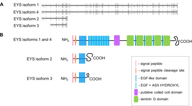 Fig. 1 EYS has four isoforms annotated in genomic and protein databases: a summary of the genomic and protein structure of EYS isoforms.1
Key structural properties of EYS:
The main role of EYS is to uphold the structure of photoreceptors, in the retina;. It also plays a part in bodily functions like cell adhesion and signal transmission, within photoreceptor cells.
| Function | Description |
| Photoreceptor Support | Maintains structural integrity of photoreceptor cells in the retina. |
| Cell Adhesion | Facilitates adhesion between photoreceptor cells and surrounding tissues. |
| Hypoxia Protection | Involved in transmitting signals within photoreceptor cells. |
| Signal Transduction | Essential for the proper development and organization of photoreceptor cells. |
EYS plays a crucial role in photoreceptor cells to preserve retinal structure while other light-sensing proteins function differently which highlights its importance in preventing photoreceptor degeneration.
1. Garcia-Delgado, Ana B., et al. "Dissecting the role of EYS in retinal degeneration: clinical and molecular aspects and its implications for future therapy." Orphanet Journal of Rare Diseases 16 (2021): 1-14. https://doi.org/10.1186/s13023-021-01843-z
The article examines the function of EYS in degeneration and delves into its molecular aspects. It presents high affinity recombinant antibodies that target EYS and have uses, in diagnosing and treating degenerative conditions.
2. Liu, Yu, et al. "Eyes shut homolog (EYS) interacts with matriglycan of O-mannosyl glycans whose deficiency results in EYS mislocalization and degeneration of photoreceptors." Scientific reports 10.1 (2020): 7795. https://doi.org/10.1038/s41598-020-64752-4
The study demonstrates that EYS binds to matriglycan structures found in O-mannosyl glycans. The absence of these glycans results in EYS misplacement which causes photoreceptor degeneration. The research demonstrates how this interaction supports retinal health and shows promise for future therapeutic applications.
3. Marques, João Pedro, et al. "EYS-associated sector retinitis pigmentosa." Graefe's Archive for Clinical and Experimental Ophthalmology (2022): 1-9. https://doi.org/10.1007/s00417-021-05411-w
The article discusses EYS-associated sector retinitis pigmentosa, highlighting EYS mutations as a cause of this retinal degeneration disorder. It explores the clinical features, genetic basis, and potential therapeutic strategies for managing this condition.
4. Otsuka, Yuki, et al. "Phototoxicity avoidance is a potential therapeutic approach for retinal dystrophy caused by EYS dysfunction." JCI insight 9.8 (2024): e174179. https://doi.org/10.1172/jci.insight.174179
The article explores phototoxicity avoidance as a potential therapeutic strategy for retinal dystrophy caused by EYS dysfunction, highlighting its mechanisms and potential benefits in managing this genetic disorder.
5. Marques, Joaõ Pedro, and Jorge Simaõ. "Double concentric hyperautofluorescent ring in EYS-associated retinitis pigmentosa." The Asia-Pacific Journal of Ophthalmology 12.1 (2023): 105. https://doi.org/10.1097/APO.0000000000000473
The article examines the double concentric hyperautofluorescent ring observed in EYS-associated retinitis pigmentosa, exploring its clinical significance and potential as a diagnostic marker for this genetic retinal disorder.
Creative Biolabs specializes in the production of high-quality EYS antibodies for research and diagnostic applications. Our portfolio includes monoclonal antibodies tailored for ELISA, Western blot, immunohistochemistry, and other research methodologies.
For further information on our EYS antibodies, custom services, or technical guidance, please reach out via info@creative-biolabs.com.
Reference
 Loading...


