GFER Antibodies
Background
The small soluble protein GFER (Growth Factor Receptor-bound Protein) exists mainly in the mitochondria and cytoplasm of cells. The protein controls mitochondrial metabolism and cellular respiration through Complex I activity regulation to maintain cellular energy homeostasis and protect cells from oxidative stress. The discovery of GFER occurred in 2003 through genomic sequencing and its high species conservation demonstrates its essential role in cellular processes. Research into metabolic diseases and cellular aging now focuses on this protein because its discovery revealed important information about mitochondrial regulation and cellular health.
Structure of GFER
The protein GFER exists as a small molecule which weighs about 14 kDa. The protein weight shows minor variations between species because of different glycosylation patterns and small amino acid sequence differences.
| Species | Human | Mouse | Rat | Yeast |
| Molecular Weight (kDa) | 14.5 | 14.3 | 14.2 | 14.0 |
| Primary Structural Differences | Composed of 125 amino acids, forms a homodimer | Shares high sequence similarity with human GFER, minor differences in non-critical regions | Contains 125 amino acids, forms a homodimer, minor sequence variations | Homologous to human GFER, essential for mitochondrial function |
The protein GFER consists of around 125 amino acids. Is a soluble protein, with a tight structure. Its unique feature lies in the formation of homodimers that're essential for its function. Unlike proteins with a heme group attached to them GFER plays a role in regulating metabolism by interacting with Complex I. Its involvement in respiration underscores its significance, in maintaining energy balance. The secondary structure of GFER comprises mostly alpha helices and beta sheets which play a role in maintaining its stability. The preserved cysteine residues found in GFER play a role in both its dimer formation and overall function.
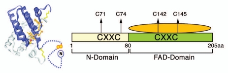 Fig. 1 Schematic model of GFER structure.1
Key structural properties of GFER:
- Forms homodimers essential for function
- Interacts with mitochondrial Complex
- Exhibits a compact structure with alpha-helices and beta-sheets
- Features conserved cysteine residues for dimerization
Functions of GFER
GFER's primary function is regulating mitochondrial metabolism and cellular respiration. However, it is also involved in various physiological processes, including cell growth regulation and protection against oxidative stress.
| Function | Description |
| Mitochondrial Metabolism Regulation | GFER modulates the activity of mitochondrial Complex I, supporting cellular respiration and energy production. |
| Cell Growth Regulation | GFER influences cell proliferation and differentiation, particularly in tissues with high metabolic demands. |
| Oxidative Stress Protection | GFER helps protect cells from oxidative damage by maintaining mitochondrial integrity and function. |
| Liver Regeneration | GFER promotes liver regeneration and tissue repair, highlighting its role in organ homeostasis. |
| Metabolic Homeostasis | GFER contributes to overall metabolic balance by regulating mitochondrial function and energy metabolism. |
The protein GFER requires homodimer formation to function whereas most mitochondrial proteins exist as monomers which demonstrates its distinct role in mitochondrial metabolic regulation.
Applications of GFER and GFER Antibody in Literature
1. Sankar, Uma, and Anthony R. Means. "Gfer is a critical regulator of HSC proliferation." Cell Cycle 10.14 (2011): 2263-2268. https://doi.org/10.4161/cc.10.14.15919
The article highlights GFER as a critical regulator of hematopoietic stem cell (HSC) proliferation, introducing its role in maintaining HSC quiescence through interaction with key cell cycle proteins. It presents evidence that GFER counteracts the activity of Jun activation domain-binding protein 1 (Jab1) by stabilizing p27kip1, thus preserving HSC functionality. This study underscores GFER's potential as a therapeutic target for diseases related to HSC dysfunction.
2. Teng, Ellen C., et al. "Gfer inhibits Jab1-mediated degradation of p27kip1 to restrict proliferation of hematopoietic stem cells." Molecular biology of the cell 22.8 (2011): 1312-1320. https://doi.org/10.1091/mbc.E10-08-0723
The research demonstrates GFER functions as a vital regulator which controls hematopoietic stem cell (HSC) proliferation while maintaining HSC quiescence through interactions with essential cell cycle proteins. The research demonstrates that GFER works against Jun activation domain-binding protein 1 (Jab1) by stabilizing p27kip1 which helps maintain HSC functionality. The research demonstrates GFER's value as a therapeutic target for diseases that result from HSC dysfunction.
Creative Biolabs: GFER Antibodies for Research
Creative Biolabs specializes in the production of high-quality GFER antibodies for research and diagnostic applications. Our portfolio includes monoclonal and polyclonal antibodies tailored for ELISA, Flow Cytometry, Western blot, immunohistochemistry, and other diagnostic methodologies.
- Custom GFER Antibody Development: We offer bespoke solutions to develop GFER antibodies tailored to your specific research needs.
- Bulk Production: We provide large-scale manufacturing of GFER antibodies to meet the demands of industry partners.
- Technical Support: Our team of experts offers consultation for optimizing protocols and troubleshooting any issues that may arise.
- Aliquoting Services: We provide GFER antibodies in conveniently sized aliquots to ensure long-term stability and consistent results in your experiments.
For additional information regarding our GFER antibodies, custom orders, or technical assistance, please reach out to us via info@creative-biolabs.com.
Reference
- Sankar, Uma, and Anthony R. Means. "Gfer is a critical regulator of HSC proliferation." Cell Cycle 10.14 (2011): 2263-2268. https://doi.org/10.4161/cc.10.14.15919
Anti-GFER antibodies
 Loading...


Hot products 
-
Mouse Anti-FLT1 Recombinant Antibody (11) (CBMAB-V0154-LY)

-
Mouse Anti-C1QC Recombinant Antibody (CBFYC-0600) (CBMAB-C0654-FY)

-
Mouse Anti-AGO2 Recombinant Antibody (V2-634169) (CBMAB-AP203LY)

-
Mouse Anti-ASTN1 Recombinant Antibody (H-9) (CBMAB-1154-CN)

-
Mouse Anti-AAV9 Recombinant Antibody (V2-634029) (CBMAB-AP023LY)

-
Mouse Anti-ANXA7 Recombinant Antibody (A-1) (CBMAB-A2941-YC)

-
Mouse Anti-BACE1 Recombinant Antibody (CBLNB-121) (CBMAB-1180-CN)

-
Mouse Anti-CFL1 Recombinant Antibody (CBFYC-1771) (CBMAB-C1833-FY)

-
Mouse Anti-CD63 Recombinant Antibody (CBXC-1200) (CBMAB-C1467-CQ)

-
Mouse Anti-BAD (Phospho-Ser136) Recombinant Antibody (CBYY-0138) (CBMAB-0139-YY)

-
Mouse Anti-AQP2 Recombinant Antibody (G-3) (CBMAB-A3359-YC)

-
Mouse Anti-CASP8 Recombinant Antibody (CBYY-C0987) (CBMAB-C2424-YY)

-
Mouse Anti-CTCF Recombinant Antibody (CBFYC-2371) (CBMAB-C2443-FY)

-
Mouse Anti-ALB Recombinant Antibody (V2-363290) (CBMAB-S0173-CQ)

-
Mouse Anti-ACO2 Recombinant Antibody (V2-179329) (CBMAB-A0627-YC)

-
Mouse Anti-C4B Recombinant Antibody (CBYY-C2996) (CBMAB-C4439-YY)

-
Mouse Anti-AHCYL1 Recombinant Antibody (V2-180270) (CBMAB-A1703-YC)

-
Mouse Anti-FAS2 Monoclonal Antibody (1D4) (CBMAB-0071-CN)

-
Rat Anti-4-1BB Recombinant Antibody (V2-1558) (CBMAB-0953-LY)

-
Mouse Anti-ARHGAP5 Recombinant Antibody (54/P190-B) (CBMAB-P0070-YC)

- AActivation
- AGAgonist
- APApoptosis
- BBlocking
- BABioassay
- BIBioimaging
- CImmunohistochemistry-Frozen Sections
- CIChromatin Immunoprecipitation
- CTCytotoxicity
- CSCostimulation
- DDepletion
- DBDot Blot
- EELISA
- ECELISA(Cap)
- EDELISA(Det)
- ESELISpot
- EMElectron Microscopy
- FFlow Cytometry
- FNFunction Assay
- GSGel Supershift
- IInhibition
- IAEnzyme Immunoassay
- ICImmunocytochemistry
- IDImmunodiffusion
- IEImmunoelectrophoresis
- IFImmunofluorescence
- IGImmunochromatography
- IHImmunohistochemistry
- IMImmunomicroscopy
- IOImmunoassay
- IPImmunoprecipitation
- ISIntracellular Staining for Flow Cytometry
- LALuminex Assay
- LFLateral Flow Immunoassay
- MMicroarray
- MCMass Cytometry/CyTOF
- MDMeDIP
- MSElectrophoretic Mobility Shift Assay
- NNeutralization
- PImmunohistologyp-Paraffin Sections
- PAPeptide Array
- PEPeptide ELISA
- PLProximity Ligation Assay
- RRadioimmunoassay
- SStimulation
- SESandwich ELISA
- SHIn situ hybridization
- TCTissue Culture
- WBWestern Blot
