IRF5 Antibodies
Background
The IRF5 gene encodes a transcription factor belonging to the interferon regulatory factor (IRF) family and is mainly expressed in immune cells. This protein plays a core role in innate immune responses and inflammatory reactions by binding to specific DNA sequences to regulate the expression of interferons and inflammation-related genes. Studies have shown that abnormal activity of IRF5 is closely related to the pathogenesis of various autoimmune diseases, such as systemic lupus erythematosus and rheumatoid arthritis. Since its functions were gradually clarified, IRF5 has become a key molecule in the study of immune signaling pathways. The molecular mechanisms of its expression regulation and activation have been widely explored, significantly enhancing people's understanding of immune regulation, cell signal transduction and autoimmune pathology.
Structure of IRF5
IRF5 is a transcription factor with a molecular weight of approximately 55 kDa. Its specific molecular weight may vary slightly due to subtypes, post-translational modifications, or species.
| Species | Human | Mouse | Rat |
| Molecular Weight (kDa) | ~55 | ~54 | ~55 |
| Primary Structural Differences | Regulating the expression of type I interferons and pro-inflammatory factors is strongly associated with autoimmune diseases | Similar functions are commonly used disease models | Commonly used in basic immunology research |
The core domain of the IRF5 protein includes the N-terminal DNA-binding domain (DBD, characterized by the tryptophan cluster), the C-terminal IRF-associated domain (IAD), and the nuclear localization signal that regulates activation. Its DBD binds to the interferon-stimulated response element (ISRE) of the target gene promoter, while IAD mediates protein-protein interactions. The key serine/threonine phosphorylation sites (such as the Ser/Thr cluster) are the core switches that activate its transcriptional activity, determining its translocation to the nucleus and its binding ability to cofactors (such as NF-κB). The activation of this protein closely regulates the polarization of immune cells and inflammatory responses.
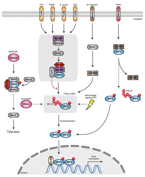 Fig. 1 A schematic representation of the proposed mechanisms of IRF5 activation.1
Key structural properties of IRF5:
- Contains N end DNA-binding domain (DBD) and C end IRF associated domain (IAD) at the core of the modularized structure
- Tryptophan pentameric clusters in DBD mediate binding to DNA specific sequences (ISRE)
- Nuclear localization signals (NLS) located in the middle of proteins regulate their nuclear transport
- Multiple serine/threonine phosphorylation sites at the C-terminal serve as key activity regulatory switches
Functions of IRF5
The IRF5 gene plays a crucial regulatory role in both innate and adaptive immune responses. Its main functions are as follows:
| Function | Description |
| Regulation of immune response | As a transcription factor, it activates the gene expression of type I interferons (such as IFN-α/β) and pro-inflammatory cytokines (such as TNF-α, IL-6, IL-12). |
| Macrophage polarization | Drive macrophages to differentiate into pro-inflammatory M1 phenotypes and enhance their antibacterial and inflammatory response capabilities. |
| Autoimmune association | Its functional abnormalities or gene polymorphisms are significantly associated with the risk of various autoimmune diseases such as systemic lupus erythematosus and rheumatoid arthritis. |
| Antiviral defense | After viral infection, it is activated by signaling pathways such as TLR, upregulates interferon expression, and establishes an antiviral state of cells. |
| Regulation of apoptosis | Under certain circumstances, it can participate in regulating the apoptosis process of immune cells and affect immune homeostasis. |
The activity of IRF5 is precisely regulated by its subcellular localization, post-translational modifications (especially phosphorylation), and protein-protein interactions. The functional imbalance of IRF5 is one of the key factors leading to immunopathology.
Applications of IRF5 and IRF5 Antibody in Literature
1. Ban, Tatsuma, et al. "Genetic and chemical inhibition of IRF5 suppresses pre-existing mouse lupus-like disease." Nature communications 12.1 (2021): 4379. https://doi.org/10.1038/s41467-021-24609-4
This study reveals that IRF5 remains abnormally activated continuously during both the active and remission phases of lupus. In model mice, partial inhibition of IRF5 is more effective in controlling the disease than complete blocking of type I interferon signals. Moreover, post-onset intervention can still inhibit disease progression and maintain remission, suggesting that IRF5 inhibitors may break through the current limitations of lupus treatment.
2. Almuttaqi, Hannah, and Irina A. Udalova. "Advances and challenges in targeting IRF5, a key regulator of inflammation." The FEBS journal 286.9 (2019): 1624-1637. https://doi.org/10.1111/febs.14654
This study reveals that IRF5 is a key regulatory factor in inflammatory responses and is involved in the pathogenesis of various autoimmune diseases such as rheumatoid arthritis and lupus. It has attracted much attention as an emerging therapeutic target. This article reviews the latest strategies targeting the IRF5 activation pathway and their therapeutic potential, and discusses the related challenges.
3. Wang, Yidong, et al. "IRF5 governs macrophage adventitial infiltration to fuel abdominal aortic aneurysm formation." JCI insight 9.3 (2024): e171488. https://doi.org/10.1172/jci.insight.171488
This study reveals that IRF5 in macrophages promotes the migration of PI3Kγ to the aortic epicendium by directly regulating it, driving the formation of abdominal aortic aneurysms (AAA). Both IRF5 and PI3Kγ are highly expressed in patient samples. Targeting this pathway can inhibit the progression of AAA.
4. Albers, G. J., et al. "IRF5 regulates airway macrophage metabolic responses." Clinical & Experimental Immunology 204.1 (2021): 134-143. https://doi.org/10.1111/cei.13573
Research has found that IRF5 is a core regulatory factor for the phenotype and metabolic response of macrophages, and can directly regulate metabolic genes such as HK2. In the influenza infection model, although the absence of IRF5 does not affect the viral load, it reduces the expression of Sirt6 and HK2 as well as their oxidative phosphorylation ability, indicating that IRF5 is crucial for the metabolic response of macrophages.
5. Wang, Zhao, et al. "An autoimmune pleiotropic SNP modulates IRF5 alternative promoter usage through ZBTB3-mediated chromatin looping." Nature Communications 14.1 (2023): 1208. https://doi.org/10.1038/s41467-023-36897-z
Research has found that the shared genetic effects of autoimmune diseases mostly originate from regulatory sequences. The key variant rs4728142 activates the short transcript of IRF5 in an allele-specific manner through the chromatin loop mediated by ZBTB3, leading to overactivation of IRF5 and polarization of M1 macrophages, revealing the fine molecular mechanism of the disease-sharing site.
Creative Biolabs: IRF5 Antibodies for Research
Creative Biolabs specializes in the production of high-quality IRF5 antibodies for research and industrial applications. Our portfolio includes monoclonal antibodies tailored for ELISA, Flow Cytometry, Western blot, immunohistochemistry, and other diagnostic methodologies.
- Custom IRF5 Antibody Development: Tailor-made solutions to meet specific research requirements.
- Bulk Production: Large-scale antibody manufacturing for industry partners.
- Technical Support: Expert consultation for protocol optimization and troubleshooting.
- Aliquoting Services: Conveniently sized aliquots for long-term storage and consistent experimental outcomes.
For more details on our IRF5 antibodies, custom preparations, or technical support, contact us at email.
Reference
- Almuttaqi, Hannah, and Irina A. Udalova. "Advances and challenges in targeting IRF5, a key regulator of inflammation." The FEBS journal 286.9 (2019): 1624-1637. https://doi.org/10.1111/febs.14654
Anti-IRF5 antibodies
 Loading...


Hot products 
-
Mouse Anti-BACE1 Recombinant Antibody (61-3E7) (CBMAB-1183-CN)

-
Mouse Anti-FPR2 Recombinant Antibody (1D6) (CBMAB-F2628-CQ)

-
Mouse Anti-CD24 Recombinant Antibody (ALB9) (CBMAB-0176CQ)

-
Rat Anti-CD300A Recombinant Antibody (172224) (CBMAB-C0423-LY)

-
Mouse Anti-Acetyl SMC3 (K105/K106) Recombinant Antibody (V2-634053) (CBMAB-AP052LY)

-
Mouse Anti-CCDC6 Recombinant Antibody (CBXC-0106) (CBMAB-C5397-CQ)

-
Mouse Anti-AFDN Recombinant Antibody (V2-58751) (CBMAB-L0408-YJ)

-
Mouse Anti-FN1 Monoclonal Antibody (D6) (CBMAB-1240CQ)

-
Mouse Anti-DMD Recombinant Antibody (D1190) (CBMAB-D1190-YC)

-
Rat Anti-4-1BB Recombinant Antibody (V2-1558) (CBMAB-0953-LY)

-
Mouse Anti-ADGRE2 Recombinant Antibody (V2-261270) (CBMAB-C0813-LY)

-
Rat Anti-AChR Recombinant Antibody (V2-12500) (CBMAB-0990-CN)

-
Mouse Anti-8-oxoguanine Recombinant Antibody (V2-7719) (CBMAB-1898CQ)

-
Mouse Anti-ARHGAP5 Recombinant Antibody (54/P190-B) (CBMAB-P0070-YC)

-
Mouse Anti-DES Monoclonal Antibody (440) (CBMAB-AP1857LY)

-
Mouse Anti-BSN Recombinant Antibody (219E1) (CBMAB-1228-CN)

-
Rabbit Anti-AP2M1 (Phosphorylated T156) Recombinant Antibody (D4F3) (PTM-CBMAB-0610LY)

-
Mouse Anti-GFAP Recombinant Antibody (24) (CBMAB-G2927-LY)

-
Mouse Anti-ARIH1 Recombinant Antibody (C-7) (CBMAB-A3563-YC)

-
Rabbit Anti-Acetyl-Histone H3 (Lys36) Recombinant Antibody (V2-623395) (CBMAB-CP0994-LY)

- AActivation
- AGAgonist
- APApoptosis
- BBlocking
- BABioassay
- BIBioimaging
- CImmunohistochemistry-Frozen Sections
- CIChromatin Immunoprecipitation
- CTCytotoxicity
- CSCostimulation
- DDepletion
- DBDot Blot
- EELISA
- ECELISA(Cap)
- EDELISA(Det)
- ESELISpot
- EMElectron Microscopy
- FFlow Cytometry
- FNFunction Assay
- GSGel Supershift
- IInhibition
- IAEnzyme Immunoassay
- ICImmunocytochemistry
- IDImmunodiffusion
- IEImmunoelectrophoresis
- IFImmunofluorescence
- IGImmunochromatography
- IHImmunohistochemistry
- IMImmunomicroscopy
- IOImmunoassay
- IPImmunoprecipitation
- ISIntracellular Staining for Flow Cytometry
- LALuminex Assay
- LFLateral Flow Immunoassay
- MMicroarray
- MCMass Cytometry/CyTOF
- MDMeDIP
- MSElectrophoretic Mobility Shift Assay
- NNeutralization
- PImmunohistologyp-Paraffin Sections
- PAPeptide Array
- PEPeptide ELISA
- PLProximity Ligation Assay
- RRadioimmunoassay
- SStimulation
- SESandwich ELISA
- SHIn situ hybridization
- TCTissue Culture
- WBWestern Blot
