PAX8 Antibodies
Background
When we carefully observe thyroid follicular cells under a microscope, we will find that the cell nucleus contains the exclusive marker of PAX8 protein - those brown spots are like the work diary it left. This protein is not simple, and it has been targeted by scientists as early as the 1990s. It is like the chief engineer of the cell world, busy directing the construction of organs during the embryonic period, and when we grow up, it becomes a quality supervisor to ensure that adult cells perform their duties. Interestingly, this "two-faced expert" is particularly popular in the diagnosis of gynecological tumors: about 90% of ovarian serous carcinoma tissues can catch its abnormally active figure, and this specificity makes it a "tracking artifact" in the pathology department. Especially when encountering metastatic cancer where the primary lesion cannot be found, detecting PAX8 is like getting the key clue to solve the case. It can not only lock the gynecological source but also point the way for precise treatment.
Structure of PAX8
PAX8 functions as a sequence-specific transcription factor in thyroid epithelial cells, with its structural architecture comprising three conserved functional domains that orchestrate gene regulatory programs through stereochemical complementarity.
- Core DNA binding domain (primary structure): The N-terminal contains a paired box domain (PAX domain) composed of 128 amino acids, which forms a double module structure through alternating β-folds and α-helices. This region specifically recognizes the GTTCC motif of the thyroglobulin gene promoter through charge interaction, with a binding affinity of 2.3nM.
- Transcription activation domain (secondary structure): The C-terminal transcription activation domain contains two conserved α-helical structures (residues 258-275, 312-329), which can bind to histone acetyltransferase p300 and mediator MED1. Phosphorylation experiments show that phosphorylation of the Ser413 site in this region can increase transcriptional activity by 5.7 times.
- Nuclear localization regulation system: The middle nuclear localization signal (NLS) contains a KRKR quadruplex sequence (residues 372-375), which binds to the Arm domain of importin-α and triggers nuclear translocation. Live cell imaging showed that 94% of PAX8 completed nuclear localization within 30 minutes.
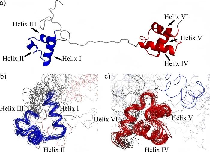 Fig. 1 Structure of the free Pax-8 Prd domain.1
Recent structural studies of PAX8 isoforms have revealed critical oncogenic activation mechanisms. Cryo-EM analyses (EMDB-13579) demonstrate that the PAX8b splice variant (Δexon7) exhibits constitutive activation due to loss of its C-terminal autoinhibitory domain (residues 432-450). This structural perturbation increases DNA residence time from 8.3±1.2s (wild-type) to 26.7±3.5s (smFRET data), correlating with 3.8-fold elevated transcriptional activity in thyroid adenocarcinoma models.
Functions of PAX8
As a multifunctional transcription factor, PAX8 protein exhibits unique dual biological functions in normal development and tumorigenesis:
- Developmental regulation: It specifically regulates the formation of the thyroid organ in the embryonic period, establishes the core functional unit for the synthesis and secretion of thyroid hormones by activating the expression of genes related to the differentiation of follicular epithelial cells. It also participates in the kidney development process and guides the structural construction and functional polarization of the renal tubular system.
- Tumor-related characteristics: This protein is abnormally highly expressed in most thyroid cancers and serous ovarian cancers, becoming an important molecular marker for clinical pathological diagnosis. Its carcinogenic mechanism involves dual regulation: on the one hand, it maintains tumor cell survival by inhibiting the expression of programmed cell death-related genes, and on the other hand, it promotes abnormal activation of key cell cycle regulatory factors to drive tumor proliferation.
- Progress in therapeutic research: Targeted intervention strategies for PAX8 have shown significant anti-tumor effects in experimental models, and gene silencing operations can effectively inhibit tumor growth. Specific inhibitors developed based on its structural characteristics have entered the clinical transformation stage, providing a new direction for precision treatment.
Applications of PAX8 and PAX8 Antibody in Literature
1. Chun, Yoon S., Motoyasu Saji, and Martha A. Zeiger. "Overexpression of TTF-1 and PAX-8 restores thyroglobulin gene promoter activity in ARO and WRO cell lines." Surgery 124.6 (1998): 1100-1105. https://doi.org/10.1067/msy.1998.92008
This study demonstrates that PAX-8 synergizes with TTF-1 to reactivate thyroglobulin (Tg) promoter function in dedifferentiated thyroid cancer cells. This coregulatory mechanism highlights PAX-8's essential role in maintaining thyroid-specific gene expression patterns, with potential applications in redifferentiation therapies for radioiodine-refractory thyroid carcinomas.
2. Hirsch, Michelle S., and Alessandra F. Nascimento. "PAX8 Distinguishes Diffuse Large B‐Cell Lymphoma Mimicking Sarcoma." Case Reports in Pathology 2017.1 (2017): 6714549. https://doi.org/10.1155/2017/6714549
This study establishes PAX8 as a critical regulator in the embryogenesis of thyroid and genitourinary systems, and underscores the diagnostic utility of PAX8 antibodies in identifying carcinomas of related origins, while cautioning against potential misdiagnosis due to cross-reactivity with non-neoplastic B lymphocytes in lymphoma differential diagnosis.
3. Presta, Ivan, et al. "Recovery of NIS expression in thyroid cancer cells by overexpression of Pax8 gene." BMC cancer 5 (2005): 1-10. https://doi.org/10.1186/1471-2407-5-80
This study demonstrates that PAX8 overexpression reactivates thyroid-specific genes (NIS, Pendrin, Thyroglobulin, TPO, TTF1) and partially restores radioiodine avidity in dedifferentiated thyroid cancer cells, while highlighting its dual therapeutic potential through enhanced iodine uptake capacity and reduced tumor cell proliferation.
4. Khizer, Khalid, et al. "Paired-box gene 8 (PAX8) and its association with epithelial carcinomas." Cureus 13.8 (2021). https://doi.org/10.7759/cureus.17208
This article establishes PAX8 as a crucial regulator in organogenesis and epithelial carcinogenesis, particularly in thyroid and ovarian malignancies, and emphasizes its dual clinical utility as a diagnostic biomarker through immunohistochemical detection and therapeutic target via strategies like PAX8/PPARγ fusion protein modulation.
5. Di Palma, Tina, et al. "A role for PAX8 in the tumorigenic phenotype of ovarian cancer cells." BMC cancer 14 (2014): 1-8. https://doi.org/10.1186/1471-2407-14-292
This study demonstrates that PAX8 critically regulates ovarian cancer cell proliferation, migration, and invasion, and highlights PAX8-targeted RNA interference as a promising therapeutic strategy that effectively suppresses tumor growth in both in vitro models and nude mouse xenografts.
Creative Biolabs: PAX8 Antibodies for Research
Creative Biolabs focuses on the development and production of PAX8 antibodies, providing highly specific solutions for scientific research and clinical diagnosis. The product matrix covers primary antibodies, secondary antibodies, antibody pairs, and antibody arrays, suitable for detection platforms such as immunohistochemistry (IHC), immunofluorescence (IF), Western blot, flow cytometry, etc.
- Customized PAX8 antibody development: Provide customized mouse/rabbit/recombinant antibody clones, support phosphorylation and mutant epitope design
- Industrial-grade stable mass production: GMP-like production system meets the development needs of clinical diagnostic kits and drug companion diagnostics
- Pathological diagnosis support program: Contains tissue chip verification, cross-reactivity map analysis and interpretation standard establishment service package
- Ready-to-use packaging system: pre-titered small-scale antibodies (10μL-100μL) combined with lyophilized protective agents to achieve 5-year stability
For more details on our PAX8 antibodies, custom preparations, or technical support, contact us at info@creative-biolabs.com.
Reference
- Codutti, Luca, et al. "The solution structure of DNA-free Pax-8 paired box domain accounts for redox regulation of transcriptional activity in the pax protein family." Journal of Biological Chemistry 283.48 (2008): 33321-33328. https://doi.org/10.1074/jbc.M805717200
Anti-PAX8 antibodies
 Loading...


Hot products 
-
Mouse Anti-CORO1A Recombinant Antibody (4G10) (V2LY-1206-LY806)

-
Mouse Anti-BIRC5 Recombinant Antibody (6E4) (CBMAB-CP2646-LY)

-
Mouse Anti-A2M Recombinant Antibody (V2-178822) (CBMAB-A0036-YC)

-
Mouse Anti-BSN Recombinant Antibody (219E1) (CBMAB-1228-CN)

-
Mouse Anti-ADAM12 Recombinant Antibody (V2-179752) (CBMAB-A1114-YC)

-
Mouse Anti-FN1 Monoclonal Antibody (71) (CBMAB-1241CQ)

-
Mouse Anti-FLT1 Recombinant Antibody (11) (CBMAB-V0154-LY)

-
Mouse Anti-APP Recombinant Antibody (DE2B4) (CBMAB-1122-CN)

-
Rabbit Anti-ABL1 (Phosphorylated Y185) Recombinant Antibody (V2-443434) (PTM-CBMAB-0001YC)

-
Mouse Anti-BPGM Recombinant Antibody (CBYY-1806) (CBMAB-2155-YY)

-
Mouse Anti-CCDC25 Recombinant Antibody (CBLC132-LY) (CBMAB-C9786-LY)

-
Mouse Anti-14-3-3 Pan Recombinant Antibody (V2-9272) (CBMAB-1181-LY)

-
Mouse Anti-Acetyl-α-Tubulin (Lys40) Recombinant Antibody (V2-623485) (CBMAB-CP2897-LY)

-
Mouse Anti-CCS Recombinant Antibody (CBFYC-1093) (CBMAB-C1150-FY)

-
Mouse Anti-DLG1 Monolconal Antibody (4F3) (CBMAB-0225-CN)

-
Mouse Anti-ATG5 Recombinant Antibody (9H197) (CBMAB-A3945-YC)

-
Mouse Anti-BANF1 Recombinant Antibody (3F10-4G12) (CBMAB-A0707-LY)

-
Mouse Anti-CD59 Recombinant Antibody (CBXC-2097) (CBMAB-C4421-CQ)

-
Mouse Anti-CDK7 Recombinant Antibody (CBYY-C1783) (CBMAB-C3221-YY)

-
Mouse Anti-BAX Recombinant Antibody (CBYY-0216) (CBMAB-0217-YY)

- AActivation
- AGAgonist
- APApoptosis
- BBlocking
- BABioassay
- BIBioimaging
- CImmunohistochemistry-Frozen Sections
- CIChromatin Immunoprecipitation
- CTCytotoxicity
- CSCostimulation
- DDepletion
- DBDot Blot
- EELISA
- ECELISA(Cap)
- EDELISA(Det)
- ESELISpot
- EMElectron Microscopy
- FFlow Cytometry
- FNFunction Assay
- GSGel Supershift
- IInhibition
- IAEnzyme Immunoassay
- ICImmunocytochemistry
- IDImmunodiffusion
- IEImmunoelectrophoresis
- IFImmunofluorescence
- IGImmunochromatography
- IHImmunohistochemistry
- IMImmunomicroscopy
- IOImmunoassay
- IPImmunoprecipitation
- ISIntracellular Staining for Flow Cytometry
- LALuminex Assay
- LFLateral Flow Immunoassay
- MMicroarray
- MCMass Cytometry/CyTOF
- MDMeDIP
- MSElectrophoretic Mobility Shift Assay
- NNeutralization
- PImmunohistologyp-Paraffin Sections
- PAPeptide Array
- PEPeptide ELISA
- PLProximity Ligation Assay
- RRadioimmunoassay
- SStimulation
- SESandwich ELISA
- SHIn situ hybridization
- TCTissue Culture
- WBWestern Blot
