TAP2 Antibodies
Background
TAP2 gene encoding the transporters in endoplasm omentum, belong to the family of ABC transporters, mainly expressed in antigen-presenting cells. This protein is responsible for transporting the antigenic peptides produced by the degradation of proteasomes in the cytoplasm to the endoplasmic reticulum lumen, where they bind to newly synthesized MHC Class I molecules, thereby initiating an adaptive immune response against intracellular pathogens. This gene was identified by multiple research teams through functional complementary cloning technology in the early 1990s. The clarification of its structure and function revealed the key mechanism of antigen processing and presentation, providing a molecular basis for understanding autoimmune diseases, infection immunity and tumor immune escape. The polymorphism of this gene directly affects the efficiency of antigen presentation and is closely related to the susceptibility to various autoimmune diseases, thus becoming an important research target in immunogenetics and clinical medicine.
Structure of TAP2
The molecular weight of the protein encoded by the TAP2 gene is approximately 75 kDa. This protein is a key component of the antigen processing-related transporter (TAP) complex, responsible for transporting antigen peptides from the cytosol to the endoplasmic reticulum lumen.
| Species | Human | Mouse | Rat |
| Molecular Weight (kDa) | About 75 | About 74.5 | About 74.8 |
| Primary Structural Differences | The typical structure of ABC transporters domain | ATP combining structural domain highly conservative | Minor interspecies variation was observed in the transmembrane helix region |
This protein is composed of approximately 700 amino acid residues, and its primary structure includes a hydrophobic transmembrane domain (TMD) and a hydrophilic nucleotide-binding domain (NBD). The core of its protein structure is a peptide binding and transport channel surrounded by multiple transmembrane α -helices. The secondary structure of this protein is mainly composed of α -helices, supplemented by connecting ring regions, which together form a "concave" structure open to the cytoplasmic side for capturing antigenic peptides. The key to its function lies in two highly conserved Walker A and Walker B motifs, which are responsible for binding and hydrolyzing ATP to provide energy for the transmembrane transport of peptides. The entire transport process strictly relies on ATP hydrolysis, ensuring the specificity and efficiency of the MHC Class I molecule antigen presentation pathway.
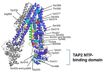 Fig. 1 The 3D structure of TAP1 (gray), TAP2 (blue), and ICP47 (green) is shown.1
Key structural properties of TAP2:
- Typical ABC transporter structure
- Hydrophobic transmembrane channels composed of multiple α-helices
- Highly conservative Walker A/Walker B motifs
- Specific peptide binding pockets
Functions of TAP2
The core function of the TAP2 gene is to transport antigen peptides from the cytoplasm to the endoplasmic reticulum lumen, and it is the rate-limiting step in the MHC Class I molecule antigen presentation pathway. Meanwhile, this gene is also involved in regulating a wider range of physiological and pathological processes such as endoplasmic reticulum stress and autophagy.
| Function | Description |
| Antigenic peptide transport | Specifically recognizing and transporting peptide segments of 8 to 16 amino acids in length, it provides an antigen source for MHC Class I molecules and is the initiating link of adaptive immune responses. |
| Immune surveillance | By maintaining the normal antigen presentation of MHC Class I molecules, cytotoxic T cells can recognize and eliminate virus-infected cells or cancerous cells. |
| Autoimmune regulation | The absence or abnormality of its function can lead to defects or alterations in the presentation of self-antigens, which is associated with susceptibility to various autoimmune diseases, such as type 1 diabetes and rheumatoid arthritis. |
| Maintenance of endoplasmic reticulum homeostasis | Load effects in the endoplasmic reticulum of peptides and circulation, involved in protein folding reactions, associated with endoplasmic reticulum stress level. |
| Tumor immune escape | Down-regulated or functionally impaired in many tumors, it is one of the important mechanisms by which tumor cells evade immune surveillance and has become a potential target for immunotherapy. |
Unlike other ABC transporters with broad substrate specificity, TAP shows a clear length preference and limited sequence tolerance for peptide substrate selectivity, which ensures the specificity and efficiency of its transport and enables it to play an irreplaceable "gatekeeper" role in the immune system.
Applications of TAP2 and TAP2 Antibody in Literature
- Ranjan, Kishu, et al. "IL-4 mediated TAP2 downregulation is a dominant and reversible mechanism of immune evasion and immunotherapy resistance in non-small cell lung cancer." Molecular Cancer 24.1 (2025): 80. https://doi.org/10.1186/s12943-025-02276-z
The article indicates that in non-small cell lung cancer, the down-regulation of TAP2 protein is associated with resistance to immunotherapy. Studies have found that the IL-4 signaling pathway can inhibit the expression of TAP2, leading to a decline in the presentation ability of tumor antigens. Targeting IL-4Rα or using epigenetic drugs to restore TAP2 function is expected to become a new strategy to overcome immune resistance.
- Cheng, Zhengyan, et al. "LMP2 and TAP2 impair tumor growth and metastasis by inhibiting Wnt/β-catenin signaling pathway and EMT in cervical cancer." BMC cancer 23.1 (2023): 1128. https://doi.org/10.1186/s12885-023-11639-y
Research has found that LMP2 and TAP2 proteins are overexpressed in cervical cancer tissues. They significantly inhibit the proliferation, migration and invasion of tumor cells by suppressing the Wnt/β-catenin signaling pathway and the epithelial-mesenchymal transition process.
- Park, Hyewon, et al. "TAP2, a peptide antagonist of Toll-like receptor 4, attenuates pain and cartilage degradation in a monoiodoacetate-induced arthritis rat model." Scientific Reports 10.1 (2020): 17451. https://doi.org/10.1038/s41598-020-74544-5
This study reveals that TAP2, as a TLR4 polypeptide antagonist, can effectively relieve arthritis pain in rats and delay cartilage damage, and its effect is superior to that of other chemical inhibitors.
- Nagy, Gregory, et al. "Identification of TAP2 protein variants resistant to inhibition by the HSV1 ICP47 protein." bioRxiv (2024). https://doi.org/10.1101/2024.11.18.624061
By screening the TAP2 protein mutation library, the study found that three specific variations such as T257I could enable TAP2 to resist the inhibition of the HSV-1 viral protein ICP47, but did not affect the inhibition of other viruses. This provides a key target for the development of new immunotherapy drugs.
- Feng, Minghao, et al. "MED12-STAT1-TAP2 axis regulates CD8+ T cell cytotoxicity and mediates immunotherapy outcome in non-small cell lung cancer." Functional & Integrative Genomics 25.1 (2025): 182. https://doi.org/10.1007/s10142-025-01690-2
Research has found that MED12 mutations in non-small cell lung cancer weaken antigen presentation function and CD8+ T cytotoxicity by inhibiting STAT1 transcription and down-regulating TAP2 expression. Therefore, the MED12 mutation can serve as an independent biomarker for predicting the efficacy of immunotherapy.
Creative Biolabs: TAP2 Antibodies for Research
Creative Biolabs specializes in the production of high-quality TAP2 antibodies for research and industrial applications. Our portfolio includes monoclonal antibodies tailored for ELISA, Flow Cytometry, Western blot, immunohistochemistry, and other diagnostic methodologies.
- Custom TAP2 Antibody Development: Tailor-made solutions to meet specific research requirements.
- Bulk Production: Large-scale antibody manufacturing for industry partners.
- Technical Support: Expert consultation for protocol optimization and troubleshooting.
- Aliquoting Services: Conveniently sized aliquots for long-term storage and consistent experimental outcomes.
For more details on our TAP2 antibodies, custom preparations, or technical support, contact us at email.
Reference
- Nagy, Gregory, et al. "Identification of TAP2 protein variants resistant to inhibition by the HSV1 ICP47 protein." bioRxiv (2024). https://doi.org/10.1101/2024.11.18.624061
Anti-TAP2 antibodies
 Loading...


Hot products 
-
Mouse Anti-AK4 Recombinant Antibody (V2-180419) (CBMAB-A1891-YC)

-
Mouse Anti-ENO1 Recombinant Antibody (8G8) (CBMAB-E1329-FY)

-
Mouse Anti-CHRNA9 Recombinant Antibody (8E4) (CBMAB-C9161-LY)

-
Mouse Anti-COL1A2 Recombinant Antibody (CF108) (V2LY-1206-LY626)

-
Mouse Anti-CORO1A Recombinant Antibody (4G10) (V2LY-1206-LY806)

-
Mouse Anti-BLK Recombinant Antibody (CBYY-0618) (CBMAB-0621-YY)

-
Mouse Anti-DISP2 Monoclonal Antibody (F66A4B1) (CBMAB-1112CQ)

-
Mouse Anti-CD33 Recombinant Antibody (P67.6) (CBMAB-C10189-LY)

-
Mouse Anti-ATP1A2 Recombinant Antibody (M7-PB-E9) (CBMAB-A4013-YC)

-
Mouse Anti-4-Hydroxynonenal Recombinant Antibody (V2-502280) (CBMAB-C1055-CN)

-
Mouse Anti-CD19 Recombinant Antibody (CBXC-1224) (CBMAB-C1491-CQ)

-
Mouse Anti-ADIPOR2 Recombinant Antibody (V2-179983) (CBMAB-A1369-YC)

-
Rat Anti-ABCC11 Recombinant Antibody (V2-179001) (CBMAB-A0236-YC)

-
Rat Anti-CD63 Recombinant Antibody (7G4.2E8) (CBMAB-C8725-LY)

-
Mouse Anti-AKT1/AKT2/AKT3 (Phosphorylated T308, T309, T305) Recombinant Antibody (V2-443454) (PTM-CBMAB-0030YC)

-
Mouse Anti-ARSA Recombinant Antibody (CBYC-A799) (CBMAB-A3679-YC)

-
Mouse Anti-ASTN1 Recombinant Antibody (H-9) (CBMAB-1154-CN)

-
Mouse Anti-GDF5 Recombinant Antibody (1F4) (CBMAB-G2740-LY)

-
Mouse Anti-CD1C Recombinant Antibody (L161) (CBMAB-C2173-CQ)

-
Mouse Anti-BrdU Recombinant Antibody (IIB5) (CBMAB-1038CQ)

- AActivation
- AGAgonist
- APApoptosis
- BBlocking
- BABioassay
- BIBioimaging
- CImmunohistochemistry-Frozen Sections
- CIChromatin Immunoprecipitation
- CTCytotoxicity
- CSCostimulation
- DDepletion
- DBDot Blot
- EELISA
- ECELISA(Cap)
- EDELISA(Det)
- ESELISpot
- EMElectron Microscopy
- FFlow Cytometry
- FNFunction Assay
- GSGel Supershift
- IInhibition
- IAEnzyme Immunoassay
- ICImmunocytochemistry
- IDImmunodiffusion
- IEImmunoelectrophoresis
- IFImmunofluorescence
- IGImmunochromatography
- IHImmunohistochemistry
- IMImmunomicroscopy
- IOImmunoassay
- IPImmunoprecipitation
- ISIntracellular Staining for Flow Cytometry
- LALuminex Assay
- LFLateral Flow Immunoassay
- MMicroarray
- MCMass Cytometry/CyTOF
- MDMeDIP
- MSElectrophoretic Mobility Shift Assay
- NNeutralization
- PImmunohistologyp-Paraffin Sections
- PAPeptide Array
- PEPeptide ELISA
- PLProximity Ligation Assay
- RRadioimmunoassay
- SStimulation
- SESandwich ELISA
- SHIn situ hybridization
- TCTissue Culture
- WBWestern Blot
