E2F1 Antibodies
Background
E2F1 is a crucial transcription factor belonging to the E2F family and plays a central role in cell cycle regulation. This protein forms heterodimers with members of the DP family and binds to the promoter regions of target genes, regulating the expression of genes related to DNA replication and cell cycle progression. During the transition of cells from the G1 phase to the S phase, the activity of E2F1 is precisely regulated by the retinoblastoma protein (RB). When RB is phosphorylated and inactivated, the released E2F1 can activate downstream target genes. Besides promoting cell proliferation, E2F1 also has a dual function of inducing cell apoptosis, and this balance is crucial for maintaining normal tissue homeostasis. Initially identified as a cellular target of the adenovirus E1A protein, the discovery of E2F1 laid the foundation for understanding the mechanism of cell cycle regulation. Its abnormal expression is closely related to the occurrence and development of various human cancers, making it an important target for tumor treatment research.
Structure of E2F1
E2F1 is a transcription factor that plays a crucial role in cell cycle regulation, with a molecular weight of approximately 47 kDa. This protein consists of 437 amino acids and contains multiple functional domains, including the DNA binding domain, dimerization domain, transcription activation domain, and RB protein binding domain. The E2F1 protein in different species is highly conserved in sequence, especially in the DNA binding domain region, which is almost completely identical. This reflects its central position in cell cycle regulation. The table below shows the basic characteristics of E2F1 protein in several representative species:
| Species | Human | Mouse | Rat | Chicken |
| Molecular Weight (kDa) | 47 | 46 | 46 | 48 |
| Primary Structural Differences | Containing a classical domain | Highly similar to others | Similar to mice | Less conservative |
The E2F1 protein recognizes specific promoter sequences through its DNA-binding domain, while its dimerization domain is responsible for forming heterodimers with DP family proteins, which is necessary for its efficient DNA binding. The transcriptional activation domain is rich in acidic amino acids and is responsible for recruiting transcriptional co-activators, while the RB binding domain mediates the interaction with the retinoblastoma protein to achieve precise regulation of the activity of this transcription factor. When the RB protein binds to the RB binding domain of E2F1, it masks its transcriptional activation domain, thereby inhibiting the expression of target genes; when the cell receives a growth signal, RB is phosphorylated and releases E2F1, exposing the transcriptional activation domain, which can then initiate the transcription of downstream genes. This ingenious structural design ensures the precise regulation of the cell cycle process.
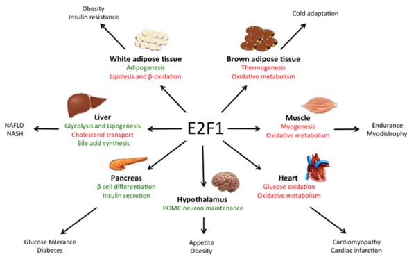 Fig. 1 Main roles of E2F1 in metabolic tissues.1
Key structural properties of E2F1:
- Contains a DNA-binding domain, responsible for recognizing specific gene sequences
- Has a dimerization domain, forming a complex with DP protein
- Abounds in transcriptional activation domains, initiating downstream gene expression
- Has an RB-binding motif, receiving cell cycle regulation signals
Functions of E2F1
The core function of E2F1 is to regulate the transcription of genes related to the cell cycle. However, it is also involved in various cellular physiological processes, including DNA damage response and apoptosis induction.
| Function | Description |
| Cell Cycle Regulation | E2F1 activates the transcription of genes necessary for the S phase, facilitating the transition of cells from the G1 phase to the S phase. |
| DNA Damage Response | When DNA is damaged, E2F1 is stabilized and participates in the repair mechanism or initiates apoptosis. |
| Apoptosis Induction | By activating p53-dependent or -independent pathways, E2F1 can lead abnormal proliferating cells towards apoptosis. |
| Differentiation Regulation | It participates in regulating the expression of differentiation-related genes in certain cell types. |
| Tumor Suppression and Promotion | Depending on the cellular environment, E2F1 can either inhibit tumor formation or promote tumor progression. |
The transcriptional activity of E2F1 is strictly regulated by the cell cycle. When it binds to the RB protein, its function is inhibited; once released from the RB inhibition, it rapidly activates the target genes. This regulatory mechanism imbalance is closely related to the occurrence of various human cancers.
Applications of E2F1 and E2F1 Antibody in Literature
1. Denechaud, Pierre-Damien, Lluis Fajas, and Albert Giralt. "E2F1, a novel regulator of metabolism." Frontiers in endocrinology 8 (2017): 311. https://doi.org/10.3389/fendo.2017.00311
The research has found that E2F1 is not only a key factor regulating cell cycle, DNA damage response and apoptosis, but also involved in metabolic regulation. The latest study has discovered that the activity of E2F1 is enhanced in obesity, which affects the overall metabolic homeostasis through tissue-specific functions, and connects the cell cycle with metabolic adaptation in cancer.
2. Fouad, Shahd, David Hauton, and Vincenzo D'Angiolella. "E2F1: cause and consequence of DNA replication stress." Frontiers in molecular biosciences 7 (2021): 599332. https://doi.org/10.3389/fmolb.2020.599332
The research has found that E2F1 regulates the cell cycle and apoptosis, and its dysfunction can lead to tumors. This article explores how abnormal signals disrupt E2F1 transcription, causing DNA replication stress, and analyzes the dual roles and post-translational regulation of E2F1 under this stress, proposing potential therapeutic targets.
3. Dubrez, Laurence. "Regulation of E2F1 transcription factor by ubiquitin conjugation." International journal of molecular sciences 18.10 (2017): 2188. https://doi.org/10.3390/ijms18102188
The research has found that the ubiquitination modification determines the stability, activity and localization of E2F1, enabling it to respond rapidly to physiological changes and stress. This article focuses on how this modification precisely regulates E2F1, a transcription factor with dual functions of promoting proliferation and inducing apoptosis.
4. Rocca, Maria Santa, et al. "E2F1 germline copy number variations and melanoma susceptibility." Journal of Translational Medicine 17.1 (2019): 181. https://doi.org/10.1186/s12967-019-1933-0
The study found that melanoma patients carry more copy number variations of the germline E2F1 gene, and heat stress can induce its expression. This indicates that both genetic and environmental factors jointly affect the susceptibility to melanoma, and E2F1 may become a potential therapeutic target.
5. Yuan, Ya-Jing, et al. "E2F1/CDK5/DRP1 axis mediates microglial mitochondrial division and autophagy in the pathogenesis of cerebral ischemia‐reperfusion injury." Clinical and Translational Medicine 15.2 (2025): e70197. https://doi.org/10.1002/ctm2.70197
The research has found that after cerebral ischemia-reperfusion injury, E2F1 activates CDK5 transcription, leading to mitochondrial division and inflammation, thereby exacerbating nerve damage. Silencing E2F1 can improve prognosis and provide a new target for the treatment of brain injury.
Creative Biolabs: E2F1 Antibodies for Research
Creative Biolabs specializes in the production of high-quality E2F1 antibodies for research and industrial applications. Our portfolio includes monoclonal and polyclonal antibodies tailored for ELISA, Flow Cytometry, Western blot, immunohistochemistry, and other diagnostic methodologies.
- Custom E2F1 Antibody Development: Tailor-made solutions to meet specific research requirements.
- Bulk Production: Large-scale antibody manufacturing for industry partners.
- Technical Support: Expert consultation for protocol optimization and troubleshooting.
- Aliquoting Services: Conveniently sized aliquots for long-term storage and consistent experimental outcomes.
For more details on our E2F1 antibodies, custom preparations, or technical support, contact us at email.
Reference
- Denechaud, Pierre-Damien, Lluis Fajas, and Albert Giralt. "E2F1, a novel regulator of metabolism." Frontiers in endocrinology 8 (2017): 311. Distributed under Open Access license CC BY 4.0, without modification. https://doi.org/10.3389/fendo.2017.00311
Anti-E2F1 antibodies
 Loading...


Hot products 
-
Mouse Anti-ADAM29 Recombinant Antibody (V2-179787) (CBMAB-A1149-YC)

-
Mouse Anti-CALR Recombinant Antibody (CBFYC-0763) (CBMAB-C0818-FY)

-
Mouse Anti-AMIGO2 Recombinant Antibody (CBYY-C0756) (CBMAB-C2192-YY)

-
Mouse Anti-ACE2 Recombinant Antibody (V2-179293) (CBMAB-A0566-YC)

-
Mouse Anti-HTLV-1 gp46 Recombinant Antibody (CBMW-H1006) (CBMAB-V208-1154-FY)

-
Mouse Anti-8-oxoguanine Recombinant Antibody (V2-7719) (CBMAB-1898CQ)

-
Rabbit Anti-CAMK2A Recombinant Antibody (BA0032) (CBMAB-0137CQ)

-
Mouse Anti-BRCA2 Recombinant Antibody (CBYY-0790) (CBMAB-0793-YY)

-
Mouse Anti-AAV9 Recombinant Antibody (V2-634029) (CBMAB-AP023LY)

-
Mouse Anti-CORO1A Recombinant Antibody (4G10) (V2LY-1206-LY806)

-
Mouse Anti-CFL1 (Phospho-Ser3) Recombinant Antibody (CBFYC-1770) (CBMAB-C1832-FY)

-
Mouse Anti-ADGRL2 Recombinant Antibody (V2-58519) (CBMAB-L0166-YJ)

-
Mouse Anti-BCL6 Recombinant Antibody (CBYY-0435) (CBMAB-0437-YY)

-
Mouse Anti-AKR1B1 Antibody (V2-2449) (CBMAB-1001CQ)

-
Mouse Anti-ATM Recombinant Antibody (2C1) (CBMAB-A3970-YC)

-
Mouse Anti-ALB Recombinant Antibody (V2-55272) (CBMAB-H0819-FY)

-
Mouse Anti-ALB Recombinant Antibody (V2-180650) (CBMAB-A2186-YC)

-
Mouse Anti-BIRC3 Recombinant Antibody (16E63) (CBMAB-C3367-LY)

-
Mouse Anti-CTNND1 Recombinant Antibody (CBFYC-2414) (CBMAB-C2487-FY)

-
Mouse Anti-BBS2 Recombinant Antibody (CBYY-0253) (CBMAB-0254-YY)

- AActivation
- AGAgonist
- APApoptosis
- BBlocking
- BABioassay
- BIBioimaging
- CImmunohistochemistry-Frozen Sections
- CIChromatin Immunoprecipitation
- CTCytotoxicity
- CSCostimulation
- DDepletion
- DBDot Blot
- EELISA
- ECELISA(Cap)
- EDELISA(Det)
- ESELISpot
- EMElectron Microscopy
- FFlow Cytometry
- FNFunction Assay
- GSGel Supershift
- IInhibition
- IAEnzyme Immunoassay
- ICImmunocytochemistry
- IDImmunodiffusion
- IEImmunoelectrophoresis
- IFImmunofluorescence
- IGImmunochromatography
- IHImmunohistochemistry
- IMImmunomicroscopy
- IOImmunoassay
- IPImmunoprecipitation
- ISIntracellular Staining for Flow Cytometry
- LALuminex Assay
- LFLateral Flow Immunoassay
- MMicroarray
- MCMass Cytometry/CyTOF
- MDMeDIP
- MSElectrophoretic Mobility Shift Assay
- NNeutralization
- PImmunohistologyp-Paraffin Sections
- PAPeptide Array
- PEPeptide ELISA
- PLProximity Ligation Assay
- RRadioimmunoassay
- SStimulation
- SESandwich ELISA
- SHIn situ hybridization
- TCTissue Culture
- WBWestern Blot
