TK1 Antibodies
Background
TK1 (thymidine kinase 1) is a key enzyme existing in the cytoplasm. It catalyzes the phosphorylation of thymidine-to-thymidine monophosphate (TMP) in the salvage pathway of DNA synthesis. Its expression level is closely related to cell proliferation, especially with the highest activity during the S phase of the cell cycle. As an important biomarker of cell proliferation, TK1 is often significantly elevated in malignant tumor tissues, and thus has significant value in cancer diagnosis and therapeutic effect monitoring. This enzyme was discovered in the mid-20th century. Its strict dependence on the cell cycle (different from mitochondrial TK2) and its key role in DNA replication have made it a key target in tumor treatment research. With the deepening of research, the relationship between the structure and function of TK1 has been continuously clarified, which not only promotes the development of targeted therapy and diagnostic technologies, but also shows broad clinical application prospects in the fields of oncology and virology, especially playing an irreplaceable role in disease progression assessment and treatment effect monitoring.
Structure of TK1
TK1 is a key metabolic enzyme with a molecular weight of approximately 25kDa (in monomer form), and its molecular weight varies slightly among different species. The following is a comparison table of molecular weights of the main species:
| Species | Human | Mouse | Bovine | Rat |
| 25.0 | 25.0 | 24.8 | 25.2 | 24.9 |
| Primary Structural Differences | Conserved catalytic domain | There are subtle variations in the N-terminal region | Dimer form more stable | Highly similar to human TK1 |
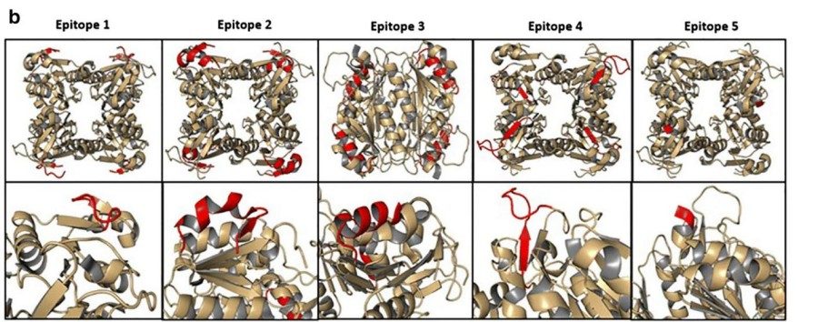
The tertiary structure of TK1 is mainly maintained stable through α -helix and β -fold, and this special spatial conformation forms a fully functional active center. This active center is highly selective and can precisely recognize and bind thymidine and ATP molecules. Research has found that the lysine residue at position 13 of the human TK1 protein (Lys13) is particularly crucial as it directly participates in the molecular process of substrate recognition. In addition, the normal functioning of enzyme activity also depends on the auxiliary effect of divalent metal ions such as Mg²⁺or Mn²⁺. It is worth noting that, unlike TK2 in mitochondria, the expression level of TK1 changes significantly along with the progress of the cell cycle, especially during the S phase when DNA synthesis is most active, when its expression reaches its peak.
 Fig. 1 Three-dimensional structure model of TK1 tetramer based on PyMOL.1
Key structural properties of TK1:
- Unique α/β folded domains constitute the active centers
- Conserved nucleotidine-binding pocket (Walker A motif)
- Specifically recognize the catalytic domain of thymidine
- Phosphate transfer mechanism dependent on divalent metal ions (Mg²⁺/Mn²⁺)
Functions of TK1
The core function of TK1 is to participate in the remedial pathways of DNA synthesis, and it also plays an important role in cell cycle regulation and disease diagnosis. Its main functions are as follows.
| Function | Description |
| DNA precursor synthesis | Catalytic thymidine phosphorylation to thymidine monophosphate (TMP) provides essential raw materials for DNA replication. |
| Cell cycle regulation | Expression level by cell cycle regulation strictly, S period activity, is the key to cell proliferation. |
| Tumor marker | Abnormally high expression in malignant tumors, has become a variety of clinical diagnosis index of cancer. |
| Antiviral target | As a key enzyme for the replication of DNA viruses such as herpes virus, it is an important target for antiviral drugs. |
| Therapeutic monitoring | Serum TK1 activity change can reflect the tumor treatment and relapse. |
The enzymatic kinetics characteristics of TK1 are characterized by a typical Michaelis curve, and its catalytic efficiency (kcat/Km) is approximately 1.5×10⁵ M-1s-1, indicating its high specificity for thymidine substrates. This characteristic makes it an ideal diagnostic marker and therapeutic target.
Applications of TK1 and TK1 Antibody in Literature
1. Wang, Naining, et al. "Investigation on cell proliferation with a new antibody against thymidine kinase 1." Analytical Cellular Pathology 23.1 (2001): 11-19. https://doi.org/10.1155/2001/658312
This article introduces that thymidine kinase 1 (TK1) is a key enzyme in DNA synthesis, and its activity is closely related to cell proliferation. The C-terminal polyclonal antibody developed in this study confirmed that TK1 was located in the cytoplasm, and its expression level increased threes from the late G1 to the G2 phase, making it a specific biomarker for cell proliferation. This antibody is suitable for the detection of archived specimens and shows good specificity in both normal and malignant proliferating tissues.
2. Jagarlamudi, K. K., Venge P, and Eriksson S. "Analytical and clinical characterization of an optimized dual monoclonal sandwich ELISA for the quantification of thymidine kinase 1 (TK1) protein in human blood samples." PLoS One 17.10 (2022): e0275444. https://doi.org/10.1371/journal.pone.0275444
This article indicates that TK1 is a key enzyme in DNA synthesis, and its activity can be used as a biomarker for monitoring the prognosis and therapeutic effect of malignant tumors. The TK 210 ELISA kit developed by AroCell Company uses double monoclonal antibody technology to detect TK1 protein. Methodological validation shows high specificity, good precision, and sample stability. Clinical verification shows that in hematological malignancies (n=100), breast cancer (n=56), and prostate cancer (n=70), the sensitivity of this kit is comparable to that of the gold standard [³H]-dThd phosphorylation method or even better. The specificity reaches 98%. Studies have confirmed that TK 210 ELISA is an accurate and reliable tool for TK1 protein detection, which can enhance the clinical application value of TK1 in cancer management.
3. Bitter, Eliza E., et al. "Thymidine kinase 1 through the ages: a comprehensive review." Cell & bioscience 10 (2020): 1-16. https://doi.org/10.1186/s13578-020-00493-1
This article indicates that TK1 is a key enzyme for cell proliferation and is involved in the DNA salvage synthesis pathway. The upregulation of its expression is an early event in cancer. The level of serum TK1 is associated with tumor stage and poor prognosis, and can more accurately reflect the rate of tumor DNA synthesis. Compared with proliferation markers such as PCNA and Ki-67, TK1 has more advantages in cancer recurrence monitoring, therapeutic effect evaluation and survival prediction. This article reviews the regulatory mechanism of TK1, its role in tumorigenesis, and its potential as a biomarker and therapeutic target.
4. Velazquez, Edwin J., et al. "Selection of human single domain antibodies (sdAb) against thymidine kinase 1 and their incorporation into sdAb-Fc antibody constructs for potential use in cancer therapy." Plos one 17.3 (2022): e0264822. https://doi.org/10.1371/journal.pone.0264822
This article indicates that TK1 is not only a prognostic marker for cancer but may also be a therapeutic target. In this study, for the first time, the antibody fragment specifically targeting TK1 was screened out from the human single-domain antibody (sdAb) library through phage display technology, with a detection limit of 3.9 ng/ml. These antibodies can recognize membrane-bound TK1 (mTK1) of tumor cells such as lung cancer (95%) and colon cancer (87%), and after fusing with the IgG1 Fc fragment, they can significantly induce antibody-dependent cytotoxicity (ADCC) to kill lung cancer cells (P<0.0267). This study provides a new tool for TK1-targeted therapy.
5. Kim, Yoonhee, et al. "Serum thymidine kinase 1 protein concentrations and presence of its autoantibody as biomarkers for screening dogs with malignant tumors." Journal of Veterinary Internal Medicine 38.1 (2024): 300-307. https://doi.org/10.1111/jvim.16946
This article indicates that TK1 is a key enzyme in DNA synthesis, and its activity detection is often affected by the serum environment. Compared with the traditional enzyme activity determination, the immunoassay based on antibodies can quantify the TK1 protein more accurately. This study was the first to detect the level of TK1 autoantibody in the serum of canine malignant tumors by side-stream immunochromatography (LFIA), and compared it with the existing detection methods of TK1 protein, providing a new idea for tumor diagnosis.
Creative Biolabs: TK1 Antibodies for Research
Creative Biolabs specializes in developing premium TK1 antibodies for research and clinical diagnostics. Our portfolio includes monoclonal antibodies optimized for ELISA, flow cytometry, Western blot, immunohistochemistry, and other diagnostic methodologies.
- Custom TK1 Antibody Development: Tailor-made solutions to meet specific research requirements.
- Bulk Production: Large-scale antibody manufacturing for industry partners.
- Technical Support: Expert consultation for protocol optimization and troubleshooting.
- Aliquoting Services: Conveniently sized aliquots for long-term storage and consistent experimental outcomes.
For more details on our TK1 antibodies, custom preparations, or technical support, contact us at info@creative-biolabs.com.
Reference
- Wang, Naining, et al. "Investigation on cell proliferation with a new antibody against thymidine kinase 1." Analytical Cellular Pathology 23.1 (2001): 11-19. https://doi.org/10.1155/2001/658312
Anti-TK1 antibodies
 Loading...
Hot products 
-
Mouse Anti-CCN2 Recombinant Antibody (CBFYC-2383) (CBMAB-C2456-FY)

-
Mouse Anti-ARHGDIA Recombinant Antibody (CBCNA-009) (CBMAB-R0415-CN)

-
Mouse Anti-CFL1 (Phospho-Ser3) Recombinant Antibody (CBFYC-1770) (CBMAB-C1832-FY)

-
Rat Anti-C5AR1 Recombinant Antibody (8D6) (CBMAB-C9139-LY)

-
Mouse Anti-ACTB Recombinant Antibody (V2-179553) (CBMAB-A0870-YC)

-
Mouse Anti-APOA1 Monoclonal Antibody (CBFYR0637) (CBMAB-R0637-FY)

-
Rabbit Anti-ALOX5AP Recombinant Antibody (CBXF-1219) (CBMAB-F0750-CQ)

-
Mouse Anti-DMD Recombinant Antibody (D1190) (CBMAB-D1190-YC)

-
Mouse Anti-ATM Recombinant Antibody (2C1) (CBMAB-A3970-YC)

-
Rabbit Anti-ABL1 (Phosphorylated Y245) Recombinant Antibody (V2-505716) (PTM-CBMAB-0465LY)

-
Mouse Anti-FLI1 Recombinant Antibody (CBXF-0733) (CBMAB-F0435-CQ)

-
Mouse Anti-ABCA3 Recombinant Antibody (V2-178911) (CBMAB-A0145-YC)

-
Mouse Anti-AKT1 Recombinant Antibody (V2-180546) (CBMAB-A2070-YC)

-
Mouse Anti-CD46 Recombinant Antibody (CBFYC-0076) (CBMAB-C0085-FY)

-
Human Anti-SARS-CoV-2 S1 Monoclonal Antibody (CBFYR-0120) (CBMAB-R0120-FY)

-
Mouse Anti-CD1C Recombinant Antibody (L161) (CBMAB-C2173-CQ)

-
Mouse Anti-AGO2 Recombinant Antibody (V2-634169) (CBMAB-AP203LY)

-
Mouse Anti-CA9 Recombinant Antibody (CBXC-2079) (CBMAB-C0131-CQ)

-
Mouse Anti-FOXL1 Recombinant Antibody (CBXF-0845) (CBMAB-F0462-CQ)

-
Mouse Anti-BMI1 Recombinant Antibody (CBYC-P026) (CBMAB-P0108-YC)

- AActivation
- AGAgonist
- APApoptosis
- BBlocking
- BABioassay
- BIBioimaging
- CImmunohistochemistry-Frozen Sections
- CIChromatin Immunoprecipitation
- CTCytotoxicity
- CSCostimulation
- DDepletion
- DBDot Blot
- EELISA
- ECELISA(Cap)
- EDELISA(Det)
- ESELISpot
- EMElectron Microscopy
- FFlow Cytometry
- FNFunction Assay
- GSGel Supershift
- IInhibition
- IAEnzyme Immunoassay
- ICImmunocytochemistry
- IDImmunodiffusion
- IEImmunoelectrophoresis
- IFImmunofluorescence
- IGImmunochromatography
- IHImmunohistochemistry
- IMImmunomicroscopy
- IOImmunoassay
- IPImmunoprecipitation
- ISIntracellular Staining for Flow Cytometry
- LALuminex Assay
- LFLateral Flow Immunoassay
- MMicroarray
- MCMass Cytometry/CyTOF
- MDMeDIP
- MSElectrophoretic Mobility Shift Assay
- NNeutralization
- PImmunohistologyp-Paraffin Sections
- PAPeptide Array
- PEPeptide ELISA
- PLProximity Ligation Assay
- RRadioimmunoassay
- SStimulation
- SESandwich ELISA
- SHIn situ hybridization
- TCTissue Culture
- WBWestern Blot


