CDC6 Antibodies
Background
CDC6, as a key cell cycle regulatory protein, is mainly responsible for initiating the DNA replication process. This protein ensures that the genome is replicated only once during each cell cycle by forming a replication precursor complex. This precise regulatory mechanism is crucial for maintaining genomic stability, and its dysfunction can lead to abnormal DNA replication and genomic instability, thereby promoting tumor development. Studies have found that CDC6 is significantly highly expressed in various cancer tissues and is closely related to poor patient prognosis. Besides participating in replication regulation, CDC6 also affects the epithelial-mesenchymal transition process through transcriptional regulation and participates in DNA damage response and cell senescence regulation. This protein was first identified as a cell division cycle gene in yeast in 1995, and its homologues were later discovered in higher eukaryotes, becoming an important molecule in the field of cell cycle research.
Structure of CDC6
CDC6 belongs to the AAA+ ATPase family of proteins, with a molecular weight of approximately 60-70 kDa. There are sequence differences among different species.
| Species | Humans | Mice | Tadpole | Yeast | Fruit fly |
|---|---|---|---|---|---|
| Molecular Weight (kDa) | 62.7 | 63.2 | 65.8 | 58.4 | 64.1 |
| Primary Structural Differences | Contains the classical ATPase domain | There are differences in the N-terminal extension region | Conservation of the replication initiation active site | Lower homology but functional conservation | C-terminal regulatory sequence is unique |
This protein contains approximately 560 amino acids and is mainly composed of the N-terminal ATPase domain and the C-terminal winged-helix domain. The ATPase domain is responsible for binding and hydrolyzing ATP to drive the assembly of the replication initiation complex, while the C-terminal domain mediates the interaction with DNA and replication factor ORC. CDC6 achieves ATP binding and hydrolysis through its conserved Walker A and Walker B motifs, and its conformational changes are finely regulated by phosphorylation modifications. The cell cycle-dependent phosphorylation state can alter the subcellular localization and protein stability of CDC6, thereby ensuring that DNA replication begins only once in the S phase. In some species, CDC6 also contains additional nuclear localization signal regions, participating in the regulation of its nuclear entry transport process.
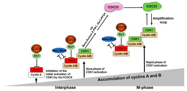 Fig. 1 A model of the role of CDC6 in the control of the M-phase entry.1
Key structural characteristics of CDC6:
- Conserved AAA+ ATPase domain, which mediates ATP binding and hydrolysis
- C-terminal winged-helix domain, involved in DNA binding and protein interaction
- Walker A and Walker B motifs, driving conformational changes to regulate replication initiation
- Multiple CDK phosphorylation sites, finely regulating protein stability and subcellular localization
Functions of CDC6
The core function of CDC6 is to initiate DNA replication and ensure that the genome is replicated only once during each cell cycle. However, it is also involved in various cellular activities, including DNA damage response, transcriptional regulation, and control of cell senescence.
| Function | Description |
|---|---|
| Initiation of replication licensing | As a key component of the pre-replication complex, it is responsible for loading the MCM helicase onto the replication origin and initiating DNA replication. |
| Maintenance of genomic stability | Ensuring that the genome is replicated only once during each cell cycle and preventing abnormal re-replication that leads to genomic damage. |
| DNA damage response | Participates in checkpoint regulation and coordinates the replication process with the repair mechanism when DNA is damaged. |
| Transcriptional regulation regulation | Combining with the promoter of target genes, it regulates the transcriptional activity of genes such as CDH1 related to epithelial-mesenchymal transition. |
| Induction of cell senescence | Regulates the senescence process through subcellular localization changes, and cytoplasmic retention can bypass the replication function and activate the senescence pathway. |
The expression and activity of CDC6 are precisely regulated by cell cycle-dependent phosphorylation, and its abnormal increase is closely related to tumor occurrence and development, reflecting the dual functional characteristics of this protein in normal physiology and pathological states.
Applications of CDC6 and CDC6 Antibody in Literature
1. El Dika, Mohammed, et al. "CDC6 as a key inhibitory regulator of CDK1 activation dynamics and the timing of mitotic entry and progression." Biology 12.6 (2023): 855. https://doi.org/10.3390/biology12060855
The article indicates that CDC6, as a regulatory factor for the S phase, cooperates with Xic1 to inhibit the activity of CDK1. This forms an independent regulatory mechanism outside the Wee1/Myt1 pathway, ensuring the precise timing of embryonic mitosis.
2. Cui, Jianfeng, et al. "Deubiquitination of CDC6 by OTUD6A promotes tumour progression and chemoresistance." Molecular Cancer 23.1 (2024): 86. https://doi.org/10.1186/s12943-024-01996-y
The study found that OTUD6A stabilizes CDC6 by removing the K6, K33, and K48 chains, and promotes the proliferation of bladder cancer and chemotherapy resistance. The two are positively correlated and predict a poor prognosis, providing a new target for cancer treatment.
3. Wang, Feng, et al. "CDC6 is a prognostic biomarker and correlated with immune infiltrates in glioma." Molecular cancer 21.1 (2022): 153. https://doi.org/10.1186/s12943-022-01623-8
The study found that CDC6 is highly expressed in glioma, and is associated with poor patient prognosis, immune infiltration, and DNA transcription. In vitro experiments confirmed that silencing CDC6 can inhibit tumor cell proliferation and migration and promote apoptosis, and it is expected to become a prognostic marker and an immunotherapy target.
4. Lim, Nicholas, and Paul A. Townsend. "Cdc6 as a novel target in cancer: oncogenic potential, senescence and subcellular localisation." International journal of cancer 147.6 (2020): 1528-1534. https://doi.org/10.1002/ijc.32900
The study found that CDC6, as a key factor in DNA replication, is highly expressed in pancreatic cancer and is associated with the KRAS pathway. It promotes EMT through transcriptional regulation and participates in the DNA damage response. Its cytoplasmic retention or induction of senescence provides new ideas for treatment.
5. Mourkioti, Ioanna, et al. "A GATA2-CDC6 axis modulates androgen receptor blockade-induced senescence in prostate cancer." Journal of Experimental & Clinical Cancer Research 42.1 (2023): 187. https://doi.org/10.1186/s13046-023-02769-z
The study found that enzalutamide induces aging of prostate cancer cells by down-regulating CDC6 through GATA2. In drug-resistant cells, the decrease in GATA2 leads to the stability of CDC6, promoting EMT and evading aging. Direct targeting of CDC6 can reverse the malignant characteristics and provide a new strategy for treating drug resistance.
Creative Biolabs: CDC6 Antibodies for Research
Creative Biolabs specializes in the production of high-quality CDC6 antibodies for research and industrial applications. Our portfolio includes monoclonal and polyclonal antibodies tailored for ELISA, Flow Cytometry, Western blot, immunohistochemistry, and other diagnostic methodologies.
- Custom CDC6 Antibody Development: Tailor-made solutions to meet specific research requirements.
- Bulk Production: Large-scale antibody manufacturing for industry partners.
- Technical Support: Expert consultation for protocol optimization and troubleshooting.
- Aliquoting Services: Conveniently sized aliquots for long-term storage and consistent experimental outcomes.
For more details on our CDC6 antibodies, custom preparations, or technical support, contact us at email.
Reference
- El Dika, Mohammed, et al. "CDC6 as a key inhibitory regulator of CDK1 activation dynamics and the timing of mitotic entry and progression." Biology 12.6 (2023): 855. Distributed under Open Access license CC BY 4.0, without modification. https://doi.org/10.3390/biology12060855
Anti-CDC6 antibodies
 Loading...


Hot products 
-
Mouse Anti-ARSA Recombinant Antibody (CBYC-A799) (CBMAB-A3679-YC)

-
Mouse Anti-ASH1L Monoclonal Antibody (ASH5H03) (CBMAB-1372-YC)

-
Mouse Anti-BBS2 Recombinant Antibody (CBYY-0253) (CBMAB-0254-YY)

-
Rabbit Anti-DLK1 Recombinant Antibody (9D8) (CBMAB-D1061-YC)

-
Mouse Anti-dsDNA Recombinant Antibody (22) (CBMAB-AP1954LY)

-
Mouse Anti-APP Recombinant Antibody (5C2A1) (CBMAB-A3314-YC)

-
Mouse Anti-CD24 Recombinant Antibody (SN3) (CBMAB-C1037-CQ)

-
Mouse Anti-ACE2 Recombinant Antibody (V2-179293) (CBMAB-A0566-YC)

-
Mouse Anti-CRTAM Recombinant Antibody (CBFYC-2235) (CBMAB-C2305-FY)

-
Rabbit Anti-BRCA2 Recombinant Antibody (D9S6V) (CBMAB-CP0017-LY)

-
Mouse Anti-GluRIIA Monoclonal Antibody (8B4D2) (CBMAB-0305-CN)

-
Mouse Anti-ABCA3 Recombinant Antibody (V2-178911) (CBMAB-A0145-YC)

-
Mouse Anti-CEMIP Recombinant Antibody (3C12) (CBMAB-K0296-LY)

-
Mouse Anti-ADIPOR2 Recombinant Antibody (V2-179983) (CBMAB-A1369-YC)

-
Mouse Anti-GIPC2 Recombinant Antibody (10) (CBMAB-G0476-LY)

-
Rat Anti-(1-5)-α-L-Arabinan Recombinant Antibody (V2-501861) (CBMAB-XB0003-YC)

-
Mouse Anti-BLK Recombinant Antibody (CBYY-0618) (CBMAB-0621-YY)

-
Rabbit Anti-CBL Recombinant Antibody (D4E10) (CBMAB-CP0149-LY)

-
Mouse Anti-GFAP Recombinant Antibody (5) (CBMAB-G0346-LY)

-
Mouse Anti-APOE Recombinant Antibody (A1) (CBMAB-0078CQ)

- AActivation
- AGAgonist
- APApoptosis
- BBlocking
- BABioassay
- BIBioimaging
- CImmunohistochemistry-Frozen Sections
- CIChromatin Immunoprecipitation
- CTCytotoxicity
- CSCostimulation
- DDepletion
- DBDot Blot
- EELISA
- ECELISA(Cap)
- EDELISA(Det)
- ESELISpot
- EMElectron Microscopy
- FFlow Cytometry
- FNFunction Assay
- GSGel Supershift
- IInhibition
- IAEnzyme Immunoassay
- ICImmunocytochemistry
- IDImmunodiffusion
- IEImmunoelectrophoresis
- IFImmunofluorescence
- IGImmunochromatography
- IHImmunohistochemistry
- IMImmunomicroscopy
- IOImmunoassay
- IPImmunoprecipitation
- ISIntracellular Staining for Flow Cytometry
- LALuminex Assay
- LFLateral Flow Immunoassay
- MMicroarray
- MCMass Cytometry/CyTOF
- MDMeDIP
- MSElectrophoretic Mobility Shift Assay
- NNeutralization
- PImmunohistologyp-Paraffin Sections
- PAPeptide Array
- PEPeptide ELISA
- PLProximity Ligation Assay
- RRadioimmunoassay
- SStimulation
- SESandwich ELISA
- SHIn situ hybridization
- TCTissue Culture
- WBWestern Blot
