MYH7 Antibodies
Background
The MYH7 gene encodes the β -myocardial myosin heavy chain, a key contractile protein mainly expressed in the heart and slow-twitch skeletal muscle. This gene product, as a core component of the myosin complex, generates mechanical force by hydrolyzing ATP, directly driving myocardial contraction and maintaining the heart's pumping function. Its mutations can lead to a variety of autosomal dominant genetic diseases, including hypertrophic cardiomyopathy and dilated cardiomyopathy. Currently, more than 400 pathogenic variations have been discovered. As the first gene confirmed to be associated with hereditary cardiomyopathy, the research on MYH7 not only revealed the connection between the structure and function of sarcomere proteins, but also promoted the application of precision medicine in the field of cardiovascular diseases, laying a molecular foundation for genetic diagnosis, risk assessment and targeted treatment strategies.
Structure of MYH7
The β -myosin heavy chain encoded by the MYH7 gene is a large protein with a molecular weight of approximately 223 kDa. There are slight differences in its molecular weight among different species, mainly due to the amino acid polymorphism of the myosin head domain.
| Species | Human | Mouse | Pig | Bovine | Zebrafish |
| Molecular Weight (kDa) | 223 | 222 | 223.5 | 223.2 | 221.8 |
| Primary Structural Differences | Containing 1935 amino acids, with ATP enzyme activity | Head structure domain has three characteristic residue replace | 95% homology with human sequence | Conservative coiled-coil tail structure | Adaptive variation exists in the motor function domain |
This protein is composed of a highly conserved N-terminal spherical head, a cervical hinge region and a C-terminal α -helical tail. Its head domain contains key actin binding sites and ATP hydrolysis active centers, and mechanical force transmission is achieved through the neck lever arm. The SH1-HELIX region encoded by exon 13 acts as a molecular switch, controlling the precision of conformational changes in the contraction cycle, while the converter domain determined by exon 19 converts chemical energy into mechanical motion.
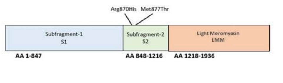 Fig. 1 MYH7 domain.1
Key structural properties of MYH7:
- Highly conserved spherical head domains
- Molecular lever mechanism for controlling the hinge area
- ATP hydrolytic activity and actin binding sites
Functions of MYH7
The MYH7 gene encodes the β -myocardial myosin heavy chain, whose core function is to act as a molecular motor for myocardial contraction. This protein is also involved in the regulation of various pathophysiological processes.
| Function | Description |
| Generation of mechanical force | Through the ATP hydrolysis activity of its head domain, it converts chemical energy into mechanical energy, directly driving the sliding and contraction of myocardial filaments. |
| Contraction regulation | Its ATPase activity is regulated by troponin-tropomyosin complex, which enables fine control of the frequency and intensity of cardiac contraction. |
| Disease occurrence | Gene mutations can disrupt its normal mechanical properties or cause misfolded proteins to accumulate in the muscle segments, leading to hypertrophic or dilated cardiomyopathy. |
| Energy steady state | As one of the most important energy-consuming proteins in myocardial cells, the alteration of its activity directly affects the energy metabolism balance of the entire cell. |
| Structural support | The α -helical coiled spiral rod-shaped structure formed at its tail provides a core structural framework and stability for the assembly of the muscle segments. |
The dynamic stroke mechanism of this protein exhibits a typical "conformation-recovery" cycle characteristic, which is in stark contrast to the synergistic effect of hemoglobin, highlighting its specificity as a molecular motor in converting nanoscale movements into macroscopic muscle contractions.
Applications of MYH7 and MYH7 Antibody in Literature
1. Wang, Lingyu, et al. "MYH7 R453C induced cardiac remodelling via activating TGF-β/Smad2/3, ERK1/2 and Nox4/ROS/NF-κB signalling pathways." Open Biology 14.6 (2024): 230427. https://doi.org/10.1098/rsob.230427
In this study, by constructing a MYH7 R453C gene mutant pig model, it was found that this mutation, by activating the TGF-β/Smad2/3, ERK1/2 and Nox4/ROS pathways, synergically induced myocardial remodels, inflammatory responses and cell death, leading to myocardial hypertrophy and fibrosis. Epigallocatechin gallate can alleviate related lesions.
2. Bollen, I. A. E., and J. van der Velden. "The contribution of mutations in MYH7 to the onset of cardiomyopathy." Netherlands Heart Journal 25.12 (2017): 653-654. https://doi.org/10.1007/s12471-017-1045-5
This Dutch study discovered a novel mutation of the MYH7 gene, p.Asn1918Lys. This mutation can lead to dilated cardiomyopathy, as well as perinatal cardiomyopathy, myocardial insufficiency and congenital heart disease, revealing the complexity of multiple cardiac phenotypes that a single MYH7 mutation can cause.
3. Friedman, Clayton E., et al. "Multiplexed functional assessments of MYH7 variants in human cardiomyocytes." Circulation: Genomic and Precision Medicine 17.2 (2024): e004377. https://doi.org/10.1161/CIRCGEN.123.004377
In this study, cardiomyocytes differentiated from stem cells were utilized to establish for the first time a high-throughput functional analysis method for missense variations of the MYH7 gene. This method can effectively distinguish pathogenic variations from benign variations by evaluating the abundance of β -myosin heavy chains and cell survival rates, and is expected to solve the clinical predicament of unclear significance of a large number of MYH7 variations.
4. Franke, Magda, et al. "A MYH7 variant in a five-generation-family with hypertrophic cardiomyopathy." Frontiers in Genetics 15 (2024): 1306333. https://doi.org/10.3389/fgene.2024.1306333
This study identified A novel MYH7 gene c.2342T>A variant in a fifth-generation HCM family. This variation leads to significant clinical heterogeneity. Among the ten carriers, eight had severe symptoms and three died suddenly, suggesting that the severity of the disease may be influenced by other genetic factors.
5. Myasnikov, Roman P., et al. "A splice variant of the MYH7 gene is causative in a family with isolated left ventricular noncompaction cardiomyopathy." Genes 13.10 (2022): 1750. https://doi.org/10.3390/genes13101750
The article indicates that traditionally, it is believed that MYH7 gene truncation variations do not cause disease. However, this study discovered a novel MYH7 splicing variation in a family with incomplete left ventricular densification. Analysis shows that not all the variants predicted to be truncated function through the haploid insufficiency mechanism, emphasizing the importance of precise assessment of MYH7 splicing variants.
Creative Biolabs: MYH7 Antibodies for Research
Creative Biolabs specializes in the production of high-quality MYH7 antibodies for research and industrial applications. Our portfolio includes monoclonal antibodies tailored for ELISA, Flow Cytometry, Western blot, immunohistochemistry, and other diagnostic methodologies.
- Custom MYH7 Antibody Development: Tailor-made solutions to meet specific research requirements.
- Bulk Production: Large-scale antibody manufacturing for industry partners.
- Technical Support: Expert consultation for protocol optimization and troubleshooting.
- Aliquoting Services: Conveniently sized aliquots for long-term storage and consistent experimental outcomes.
For more details on our MYH7 antibodies, custom preparations, or technical support, contact us at email.
Reference
- Ferradini, Valentina, et al. "Variants in MHY7 gene cause arrhythmogenic cardiomyopathy." Genes 12.6 (2021): 793. https://doi.org/10.3390/genes12060793
Anti-MYH7 antibodies
 Loading...


Hot products 
-
Mouse Anti-GGT1 Recombinant Antibody (1F9) (CBMAB-G3273-LY)

-
Mouse Anti-DMD Recombinant Antibody (D1190) (CBMAB-D1190-YC)

-
Mouse Anti-CRYAB Recombinant Antibody (A4345) (CBMAB-A4345-YC)

-
Mouse Anti-CECR2 Recombinant Antibody (CBWJC-2465) (CBMAB-C3533WJ)

-
Rat Anti-CCR2 Recombinant Antibody (475301) (CBMAB-C1338-LY)

-
Mouse Anti-ENO2 Recombinant Antibody (H14) (CBMAB-E1341-FY)

-
Mouse Anti-ABL2 Recombinant Antibody (V2-179121) (CBMAB-A0364-YC)

-
Mouse Anti-ASH1L Monoclonal Antibody (ASH5H03) (CBMAB-1372-YC)

-
Rabbit Anti-CAMK2A Recombinant Antibody (BA0032) (CBMAB-0137CQ)

-
Mouse Anti-ACO2 Recombinant Antibody (V2-179329) (CBMAB-A0627-YC)

-
Mouse Anti-CD46 Recombinant Antibody (CBFYC-0076) (CBMAB-C0085-FY)

-
Rat Anti-EPO Recombinant Antibody (16) (CBMAB-E1578-FY)

-
Mouse Anti-BIRC5 Recombinant Antibody (6E4) (CBMAB-CP2646-LY)

-
Rat Anti-ADAM10 Recombinant Antibody (V2-179741) (CBMAB-A1103-YC)

-
Mouse Anti-CCDC6 Recombinant Antibody (CBXC-0106) (CBMAB-C5397-CQ)

-
Mouse Anti-AAV9 Recombinant Antibody (V2-634029) (CBMAB-AP023LY)

-
Mouse Anti-A2M Recombinant Antibody (V2-178822) (CBMAB-A0036-YC)

-
Rabbit Anti-B2M Recombinant Antibody (CBYY-0059) (CBMAB-0059-YY)

-
Mouse Anti-DISP2 Monoclonal Antibody (F66A4B1) (CBMAB-1112CQ)

-
Rat Anti-ABCC11 Recombinant Antibody (V2-179001) (CBMAB-A0236-YC)

- AActivation
- AGAgonist
- APApoptosis
- BBlocking
- BABioassay
- BIBioimaging
- CImmunohistochemistry-Frozen Sections
- CIChromatin Immunoprecipitation
- CTCytotoxicity
- CSCostimulation
- DDepletion
- DBDot Blot
- EELISA
- ECELISA(Cap)
- EDELISA(Det)
- ESELISpot
- EMElectron Microscopy
- FFlow Cytometry
- FNFunction Assay
- GSGel Supershift
- IInhibition
- IAEnzyme Immunoassay
- ICImmunocytochemistry
- IDImmunodiffusion
- IEImmunoelectrophoresis
- IFImmunofluorescence
- IGImmunochromatography
- IHImmunohistochemistry
- IMImmunomicroscopy
- IOImmunoassay
- IPImmunoprecipitation
- ISIntracellular Staining for Flow Cytometry
- LALuminex Assay
- LFLateral Flow Immunoassay
- MMicroarray
- MCMass Cytometry/CyTOF
- MDMeDIP
- MSElectrophoretic Mobility Shift Assay
- NNeutralization
- PImmunohistologyp-Paraffin Sections
- PAPeptide Array
- PEPeptide ELISA
- PLProximity Ligation Assay
- RRadioimmunoassay
- SStimulation
- SESandwich ELISA
- SHIn situ hybridization
- TCTissue Culture
- WBWestern Blot
