S100A8 Antibodies
Background
S100A8 is a small molecule calcium-binding protein, which mainly combines with S100A9 in the form of a heterodimer to form calprotectin and is highly expressed in myeloid cells. This protein plays a key role in innate immune defense and the development of chronic inflammatory diseases by regulating inflammatory responses, cellular chemotaxis and immune response processes. Under pathological conditions, S100A8 can serve as a marker of acute inflammation, and its expression level is closely related to autoimmune diseases and the tumor microenvironment. It was first identified as a granulocyte-specific protein in 1987. The study of its three-dimensional structure revealed a unique conformational change mechanism in the EF-hand calcium-binding domain. This protein has become an important model for the study of inflammatory disease mechanisms and the development of clinical diagnostic markers, promoting a deeper understanding of the innate immune regulatory network.
Structure of S100A8
S100A8 is a small molecule protein with a molecular weight of approximately 10.8 kDa. This value shows slight differences among different species, mainly due to minor changes in amino acid sequences.
| Species | Human | Mouse | Rat | Bovine |
| Molecular Weight (kDa) | 10.8 | 10.6 | 10.7 | 10.9 |
| Primary Structural Differences | Conservative EF-hand domain | Highly homologous to humans | The key calcium binding sites are the same | The amino acid sequences are highly similar |
This protein is composed of 93 amino acids and forms a stable homodimer conformation. Its three-dimensional structure contains a pair of unique EF-hand calcium ion-binding motifs, which regulate biological activity through conformational changes. The protein surface is distributed with a large number of hydrophobic residues, mediating the formation of a heterodimer with S100A9. Two calcium-binding loops are connected through a hinge region. After calcium ion binding, conformational rearrangement occurs, thereby exposing the target protein binding site and performing its extensive intracellular and extracellular signaling functions.
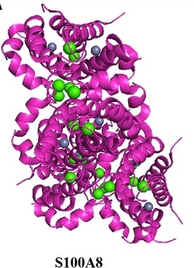 Fig. 1 Structure of S100A8.1
Key structural properties of S100A8:
- Unique EF-hand calcium ion-binding domain
- Highly conserved hydrophobic surfaces mediate dimerization
- Calcium-dependent conformational changes regulate functional activity
Functions of S100A8
The core function of the S100A8 protein is to regulate inflammatory responses and immune responses. In addition, it is also involved in pathophysiological processes such as cell migration, tumor progression and the maintenance of immune homeostasis.
| Function | Description |
| Inflammatory regulation | Released as an alarmin, it activates TLR4 and other pattern recognition receptors to initiate and amplify the innate immune response. |
| Cell chemotaxis | The formation of heterodimer calcemin with S100A9 mediates the directed migration of leukocytes to the inflammatory site. |
| Tumor regulation | High expression in tumor microenvironment, the impact of tumor cell proliferation, invasion and angiogenesis process. |
| Immune homeostasis | By regulating cytoskeletal recombination and phagocytosis, it participates in the clearance of apoptotic cells and maintains tissue homeostasis. |
| Reactive oxygen species regulation | With NADPH oxidase, regulate the reactive oxygen species (ROS) generation, affect the intracellular REDOX state. |
The heterodimer formed by S100A8 and S100A9 changes its conformation in a calcium-dependent manner. Its functional diversity stems from the different target protein binding interfaces exposed by conformational changes, thus playing multiple roles in innate immunity.
Applications of S100A8 and S100A8 Antibody in Literature
1. Wang, Yanghanzhao, et al. "S100A8/A9hi neutrophils induce mitochondrial dysfunction and PANoptosis in endothelial cells via mitochondrial complex I deficiency during sepsis." Cell Death & Disease 15.6 (2024): 462. https://doi.org/10.1038/s41419-024-06849-6
The article indicates that in sepsis, S100A8/A9 inhibits Nrf1 expression and down-regulates the I component of the mitochondrial complex Ndufa3, leading to mitochondrial dysfunction and subsequently triggering ZBp1-mediated endothelial cell PANoptosis. Clinical analysis indicates that high expression of S100A8/A9 is closely related to endothelial injury, and S100A8 is an independent risk factor for poor prognosis.
2. Ma, Jie, et al. "S100A8/A9 as a prognostic biomarker with causal effects for post-acute myocardial infarction heart failure." Nature communications 15.1 (2024): 2701. https://doi.org/10.1038/s41467-024-46973-7
This study, through a large-scale cohort, found that elevated S100A8/A9 after acute myocardial infarction is a strong predictor of heart failure, and its predictive ability is superior to traditional indicators. Mendelian randomization analysis further confirmed that S100A8/A9 is a potential pathogenic mediator leading to heart failure.
3. Sprenkeler, Evelien GG, et al. "S100A8/A9 is a marker for the release of neutrophil extracellular traps and induces neutrophil activation." Cells 11.2 (2022): 236. https://doi.org/10.3390/cells11020236
The research found that S100A8/A9 is mainly located in the cytoplasm of neutrophils rather than in the granules. Its release depends on the formation of neutrophil extracellular traps (NETs), which in turn can activate neutrophils, upregulate CD11b and promote adhesion, thereby amplifying the inflammatory response.
4. Razmkhah, Farnaz, et al. "S100A8 and S100A9 in hematologic malignancies: from development to therapy." International journal of molecular sciences 24.17 (2023): 13382. https://doi.org/10.3390/ijms241713382
This review explores the key role of S100A8 and its complexes in hematological malignancies. They influence the disease process by regulating cell phenotypes and shaping the malignant bone marrow microenvironment, and have shown important potential as diagnostic biomarkers and potential therapeutic targets.
5. Jakobsson, Gabriel, et al. "Therapeutic S100A8/A9 blockade inhibits myocardial and systemic inflammation and mitigates sepsis-induced myocardial dysfunction." Critical Care 27.1 (2023): 374. https://doi.org/10.1186/s13054-023-04652-x
The article indicates that elevated S100A8/A9 is closely associated with left ventricular dysfunction in patients with sepsis and mouse models. Studies have confirmed that blocking S100A8/A9 with the inhibitor ABR-238901 can effectively prevent and reverse cardiac dysfunction, and its effect is superior to that of dexamethasone. It is expected to become a new strategy for the treatment of septic cardiomyopathy.
Creative Biolabs: S100A8 Antibodies for Research
Creative Biolabs specializes in the production of high-quality S100A8 antibodies for research and industrial applications. Our portfolio includes monoclonal antibodies tailored for ELISA, Flow Cytometry, Western blot, immunohistochemistry, and other diagnostic methodologies.
- Custom S100A8 Antibody Development: Tailor-made solutions to meet specific research requirements.
- Bulk Production: Large-scale antibody manufacturing for industry partners.
- Technical Support: Expert consultation for protocol optimization and troubleshooting.
- Aliquoting Services: Conveniently sized aliquots for long-term storage and consistent experimental outcomes.
For more details on our S100A8 antibodies, custom preparations, or technical support, contact us at email.
Reference
- Sun, Yu, et al. "S100a8/A9 proteins: critical regulators of inflammation in cardiovascular diseases." Frontiers in Cardiovascular Medicine 11 (2024): 1394137. https://doi.org/10.3389/fcvm.2024.1394137
Anti-S100A8 antibodies
 Loading...


Hot products 
-
Mouse Anti-C5b-9 Recombinant Antibody (aE11) (CBMAB-AO138LY)

-
Mouse Anti-ASTN1 Recombinant Antibody (H-9) (CBMAB-1154-CN)

-
Mouse Anti-AAV8 Recombinant Antibody (V2-634028) (CBMAB-AP022LY)

-
Mouse Anti-BCL6 Recombinant Antibody (CBYY-0435) (CBMAB-0437-YY)

-
Mouse Anti-CD33 Recombinant Antibody (6C5/2) (CBMAB-C8126-LY)

-
Mouse Anti-DMD Recombinant Antibody (D1190) (CBMAB-D1190-YC)

-
Mouse Anti-CDK7 Recombinant Antibody (CBYY-C1783) (CBMAB-C3221-YY)

-
Human Anti-SARS-CoV-2 S1 Monoclonal Antibody (CBFYR-0120) (CBMAB-R0120-FY)

-
Mouse Anti-CCNH Recombinant Antibody (CBFYC-1054) (CBMAB-C1111-FY)

-
Mouse Anti-APP Recombinant Antibody (DE2B4) (CBMAB-1122-CN)

-
Mouse Anti-CARD11 Recombinant Antibody (CBFYC-0811) (CBMAB-C0866-FY)

-
Mouse Anti-CD24 Recombinant Antibody (SN3) (CBMAB-C1037-CQ)

-
Mouse Anti-ACTB Recombinant Antibody (V2-179553) (CBMAB-A0870-YC)

-
Mouse Anti-ACO2 Recombinant Antibody (V2-179329) (CBMAB-A0627-YC)

-
Mouse Anti-BLNK Recombinant Antibody (CBYY-0623) (CBMAB-0626-YY)

-
Mouse Anti-ACTN4 Recombinant Antibody (V2-6075) (CBMAB-0020CQ)

-
Mouse Anti-AKT1 Recombinant Antibody (V2-180546) (CBMAB-A2070-YC)

-
Mouse Anti-CD164 Recombinant Antibody (CBFYC-0077) (CBMAB-C0086-FY)

-
Rat Anti-ADAM10 Recombinant Antibody (V2-179741) (CBMAB-A1103-YC)

-
Mouse Anti-CD46 Recombinant Antibody (CBFYC-0076) (CBMAB-C0085-FY)

- AActivation
- AGAgonist
- APApoptosis
- BBlocking
- BABioassay
- BIBioimaging
- CImmunohistochemistry-Frozen Sections
- CIChromatin Immunoprecipitation
- CTCytotoxicity
- CSCostimulation
- DDepletion
- DBDot Blot
- EELISA
- ECELISA(Cap)
- EDELISA(Det)
- ESELISpot
- EMElectron Microscopy
- FFlow Cytometry
- FNFunction Assay
- GSGel Supershift
- IInhibition
- IAEnzyme Immunoassay
- ICImmunocytochemistry
- IDImmunodiffusion
- IEImmunoelectrophoresis
- IFImmunofluorescence
- IGImmunochromatography
- IHImmunohistochemistry
- IMImmunomicroscopy
- IOImmunoassay
- IPImmunoprecipitation
- ISIntracellular Staining for Flow Cytometry
- LALuminex Assay
- LFLateral Flow Immunoassay
- MMicroarray
- MCMass Cytometry/CyTOF
- MDMeDIP
- MSElectrophoretic Mobility Shift Assay
- NNeutralization
- PImmunohistologyp-Paraffin Sections
- PAPeptide Array
- PEPeptide ELISA
- PLProximity Ligation Assay
- RRadioimmunoassay
- SStimulation
- SESandwich ELISA
- SHIn situ hybridization
- TCTissue Culture
- WBWestern Blot
