
UCP1 Antibodies
Background
UCP1 is a transport protein located in the inner mitochondrial membrane and is mainly present in the brown adipose tissue of mammals. The uncoupling protein encoded by this gene uncouples oxidative phosphorylation with ATP synthesis by disrupting the proton gradient of the mitochondrial membrane, thereby converting substrate energy into thermal energy. This heat production mechanism is crucial for maintaining the body temperature balance and energy metabolism of homeothermic animals, especially playing a key role in cold adaptation and obesity regulation. This gene was first discovered in 1976. Its functional research not only revealed the molecular basis of non-shivering thermogenesis but also opened up new directions for the study of metabolic diseases and weight regulation. It has now become an important target for obesity treatment and metabolic research.
Structure of UCP1
UCP1 is a mitochondrial membrane protein with a molecular weight of approximately 32-33 kDa. There are slight differences in its precise molecular weight among different species, mainly due to the amino acid polymorphism in the gene coding region.
| Species | Human | Mouse | Rat | Hamster |
| Molecular Weight (kDa) | 32.8 | 32.6 | 32.9 | 32.7 |
| Primary Structural Differences | With six across the membrane structure field | High homology with human | The carboxyl terminal sequence is slightly different | There are specific variations in the nucleotide binding regions |
This protein is composed of approximately 306 amino acid residues, and its core structural feature is six transmembrane α -helices, which together form a central channel towards the mitochondrial matrix. The tertiary structure of proteins forms specific functional channels within the membrane, with both the carboxyl and amino ends located on the cytoplasmic side. The polar amino acid residues contained in the third transmembrane region are crucial for proton conduction, while the nucleotide binding sites located on the matrix side regulate the thermogenic activity of the protein through conformational changes.
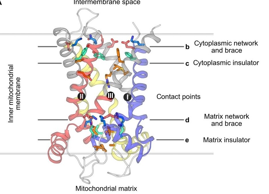 Fig. 1 Lateral view of UCP1 with the salt bridge network, braces, and insulator layers.1
Key structural properties of UCP1:
- Six transmembrane α -helical domains
- Nucleotide binding domains facing the matrix side
- Proton conduction channels formed in the third transmembrane region
Functions of UCP1
The main function of UCP1 is to mediate non-shivering thermogenesis in brown adipose tissue. In addition, this protein is also involved in a variety of metabolic regulatory processes, including energy balance regulation and adaptive thermogenesis.
| Function | Description |
| Heat generation effect | By uncoupling oxidative phosphorylation, the substrate energy is directly converted into thermal energy rather than generating ATP. |
| Energy consumption regulation | Increasing the metabolic rate and promoting the consumption of excess energy are of great significance for weight regulation. |
| Cold adaptation maintenance | Activate thermogenesis in cold environments to help warm-blooded animals maintain a stable core body temperature. |
| Metabolic protection | By reducing mitochondrial oxidative stress, it has a positive impact on metabolic health indicators such as insulin sensitivity. |
| Regulation related to obesity | The active level is associated with obesity susceptibility, activate UCP1 may against weight gain. |
The activity of UCP1 is inhibited by purine nucleotides and activated by free fatty acids. This delicate dual regulatory mechanism ensures that the heat production process is triggered only when needed, thereby achieving efficient regulation of energy metabolism.
Applications of UCP1 and UCP1 Antibody in Literature
1. Ikeda, Kenji, and Tetsuya Yamada. "UCP1 dependent and independent thermogenesis in brown and beige adipocytes." Frontiers in endocrinology 11 (2020): 498. https://doi.org/10.3389/fendo.2020.00498
The article indicates that both brown and beige adipocytes in mammals highly express UCP1 and generate heat by decoupling the mitochondrial respiratory chain. Recent studies have found that beige fats also have thermogenic pathways that do not rely on UCP1 (such as the calcium cycle mechanism). How these different mechanisms work together to regulate remains unclear, but it offers a new treatment direction for obese and diabetic patients lacking UCP1.
2. Xiong, Wei, et al. "UCP1 alleviates renal interstitial fibrosis progression through oxidative stress pathway mediated by SIRT3 protein stability." Journal of translational medicine 21.1 (2023): 521. https://doi.org/10.1186/s12967-023-04376-0
Research has found that the expression of UCP1 is significantly reduced in renal fibrosis. Enhancing UCP1 expression can inhibit oxidative stress by stabilizing SIRT3 protein, thereby reducing epithelial-mesenchymal transition and extracellular matrix accumulation, and ultimately alleviating renal fibrosis. This indicates that UCP1 is a potential new therapeutic target for chronic kidney disease.
3. Gu, Min, et al. "Decrease in UCP1 by sustained high lipid promotes NK cell necroptosis to exacerbate nonalcoholic liver fibrosis." Cell Death & Disease 15.7 (2024): 518. https://doi.org/10.1038/s41419-024-06910-4
Research has found that the absence or reduction of UCP1 in natural killer (NK) cells, in conjunction with a high-fat environment, can jointly induce mitochondrial damage and cell necrosis, thereby weakening the activity of NK cells. This functional impairment exacerbates the progression of non-alcoholic steatohepatitis to liver fibrosis.
4. Sousa-Filho, Celso Pereira Batista, and Natasa Petrovic. "No UCP1 in the kidney." Molecular Metabolism 95 (2025): 102127. https://doi.org/10.1016/j.molmet.2025.102127
In this study, through the detection of multiple specific antibodies and qPCR, it was found that the previously reported UCP1 protein signal in the kidneys still existed in UCP1 gene knockout mice, and neither the detection of highly specific antibodies nor mRNA confirmed the expression of UCP1 in the kidneys. The conclusion indicates that under strict control, UCP1 does not exist in kidney tissue.
5. Gaudry, Michael J., et al. "Terrestrial birth and body size tune UCP1 functionality in seals." Molecular biology and evolution 41.4 (2024): msae075. https://doi.org/10.1093/molbev/msae075
The study explored the evolution of UCP1 in semi-aquatic pinnipeds. The smaller Hong Kong seals have UCP1 with complete heat-producing function, which is crucial for the survival of the young on land. However, the huge elephant seals lost this function, indicating that extreme body size offset the survival advantage of UCP1-dependent thermogenesis.
Creative Biolabs: UCP1 Antibodies for Research
Creative Biolabs specializes in the production of high-quality UCP1 antibodies for research and industrial applications. Our portfolio includes monoclonal antibodies tailored for ELISA, Flow Cytometry, Western blot, immunohistochemistry, and other diagnostic methodologies.
- Custom UCP1 Antibody Development: Tailor-made solutions to meet specific research requirements.
- Bulk Production: Large-scale antibody manufacturing for industry partners.
- Technical Support: Expert consultation for protocol optimization and troubleshooting.
- Aliquoting Services: Conveniently sized aliquots for long-term storage and consistent experimental outcomes.
For more details on our UCP1 antibodies, custom preparations, or technical support, contact us at email.
Reference
- Jones, Scott A., et al. "Structural basis of purine nucleotide inhibition of human uncoupling protein 1." Science Advances 9.22 (2023): eadh4251. https://doi.org/10.1126/sciadv.adh4251
Anti-UCP1 antibodies
 Loading...
 Compare




Hot products 
-
Mouse Anti-ENO2 Recombinant Antibody (H14) (CBMAB-E1341-FY)

-
Rabbit Anti-CBL Recombinant Antibody (D4E10) (CBMAB-CP0149-LY)

-
Mouse Anti-BACE1 Recombinant Antibody (61-3E7) (CBMAB-1183-CN)

-
Mouse Anti-ALOX5 Recombinant Antibody (33) (CBMAB-1890CQ)

-
Mouse Anti-CD24 Recombinant Antibody (ALB9) (CBMAB-0176CQ)

-
Mouse Anti-ADRB2 Recombinant Antibody (V2-180026) (CBMAB-A1420-YC)

-
Mouse Anti-AAV-5 Recombinant Antibody (V2-503416) (CBMAB-V208-1402-FY)

-
Mouse Anti-APCS Recombinant Antibody (CBYC-A663) (CBMAB-A3054-YC)

-
Mouse Anti-CHRNA9 Recombinant Antibody (8E4) (CBMAB-C9161-LY)

-
Mouse Anti-AFDN Recombinant Antibody (V2-58751) (CBMAB-L0408-YJ)

-
Mouse Anti-ARSA Recombinant Antibody (CBYC-A799) (CBMAB-A3679-YC)

-
Mouse Anti-ADGRE5 Recombinant Antibody (V2-360335) (CBMAB-C2088-CQ)

-
Mouse Anti-ADGRE2 Recombinant Antibody (V2-261270) (CBMAB-C0813-LY)

-
Mouse Anti-CASQ1 Recombinant Antibody (CBFYC-0863) (CBMAB-C0918-FY)

-
Mouse Anti-GFAP Recombinant Antibody (24) (CBMAB-G2927-LY)

-
Mouse Anti-ABCA3 Recombinant Antibody (V2-178911) (CBMAB-A0145-YC)

-
Mouse Anti-AGO2 Recombinant Antibody (V2-634169) (CBMAB-AP203LY)

-
Mouse Anti-ARHGDIA Recombinant Antibody (CBCNA-009) (CBMAB-R0415-CN)

-
Mouse Anti-ELAVL4 Recombinant Antibody (6B9) (CBMAB-1132-YC)

-
Mouse Anti-CD63 Recombinant Antibody (CBXC-1200) (CBMAB-C1467-CQ)

- AActivation
- AGAgonist
- APApoptosis
- BBlocking
- BABioassay
- BIBioimaging
- CImmunohistochemistry-Frozen Sections
- CIChromatin Immunoprecipitation
- CTCytotoxicity
- CSCostimulation
- DDepletion
- DBDot Blot
- EELISA
- ECELISA(Cap)
- EDELISA(Det)
- ESELISpot
- EMElectron Microscopy
- FFlow Cytometry
- FNFunction Assay
- GSGel Supershift
- IInhibition
- IAEnzyme Immunoassay
- ICImmunocytochemistry
- IDImmunodiffusion
- IEImmunoelectrophoresis
- IFImmunofluorescence
- IGImmunochromatography
- IHImmunohistochemistry
- IMImmunomicroscopy
- IOImmunoassay
- IPImmunoprecipitation
- ISIntracellular Staining for Flow Cytometry
- LALuminex Assay
- LFLateral Flow Immunoassay
- MMicroarray
- MCMass Cytometry/CyTOF
- MDMeDIP
- MSElectrophoretic Mobility Shift Assay
- NNeutralization
- PImmunohistologyp-Paraffin Sections
- PAPeptide Array
- PEPeptide ELISA
- PLProximity Ligation Assay
- RRadioimmunoassay
- SStimulation
- SESandwich ELISA
- SHIn situ hybridization
- TCTissue Culture
- WBWestern Blot
