Antibody-Array
Antibody array (also known as antibody microarrays) is one of the most versatile approaches within multiplexed immunoassay technologies. Antibody array presents significant advantages compared with other traditional methods of protein analysis, which is widely used in a variety of research or disease diagnosis such as disease marker discovery for diagnosis, prognosis, and drug response, characterization of signaling and protein pathways, etc.
As one of the most competitive biotechnology companies in the world, Creative Biolabs has extensive expertise and experience to provide antibody array service. Our goal is to provide you with the most affordable and high-quality customer service to ensure your satisfaction in a timely and professional manner.
Introduction of Antibody Array
Antibody array is a method by immobilizing antibodies for a parallel analysis of multiple targets in a given sample. Besides common antibodies, various antibody formats have been used in arrays, such as sdAbs, single-chain variable fragments (scFvs) and fragment antigen-binding (Fab)-fragments. In addition, other technologies such as phage display and ribosome display, combined with advanced materials and bioinformatics development also facilitate the development of antibody array in recent years.
The typical workflow of an antibody array includes several steps. Simply, antibodies are primarily immobilized onto a chemically functionalized or otherwise modified surface. Once blocking the reactive groups of the surface, a sample containing targeted proteins is incubated on the array, and the proteins are captured by the antibodies. The binding results are reported directly by fluorescent labelling of the sample or by the addition of a secondary detection reagent.
Antibody arrays can be used to study a diverse number of biological processes such as protein-protein interactions, signal pathway analysis, studies of post-translation modifications, and detection of toxins. During clinical context, arrays have been used to identify novel disease biomarkers as well as generating unique proteome signature by comparing healthy and disease states. This information provides better disease management through improved diagnostics and the ability to track disease status and therapeutic efficacy.
Antibody arrays have significant advantages compared to traditional, single analyte methods of protein analysis (such as ELISA and WB). The method has become more standardized and user-friendly experimental procedures since it is high throughput, high sensitivity, and requires small sample volume. Besides, the process of antibody microarray assays is fast and takes less than 24 h from sample preparation to data interpretation compared with mainstream proteomics strategies, especially mass spectrometry (MS).
Theoretically, antibody microarrays can be designed to host a few to thousands, or even ten-thousands, of features. The array can be constructed to host multiple features for each sample, or it can be designed to divide the array into arrays that allow multiple samples to be studied at the same time. Importantly, antibody arrays can produce high dimensional data using the well-established DNA microarray software, since the images scanned from planar antibody microarrays are similar to DNA microarrays.
Antibody arrays are most often used during the biomarker discovery process. However, the cross-reactivity to non-specific proteins is still a challenge for the single binder version of the assay. Thus, uncontrolled reactions with other target proteins display one of the main obstacles in establishing high performance and high specificity assays.
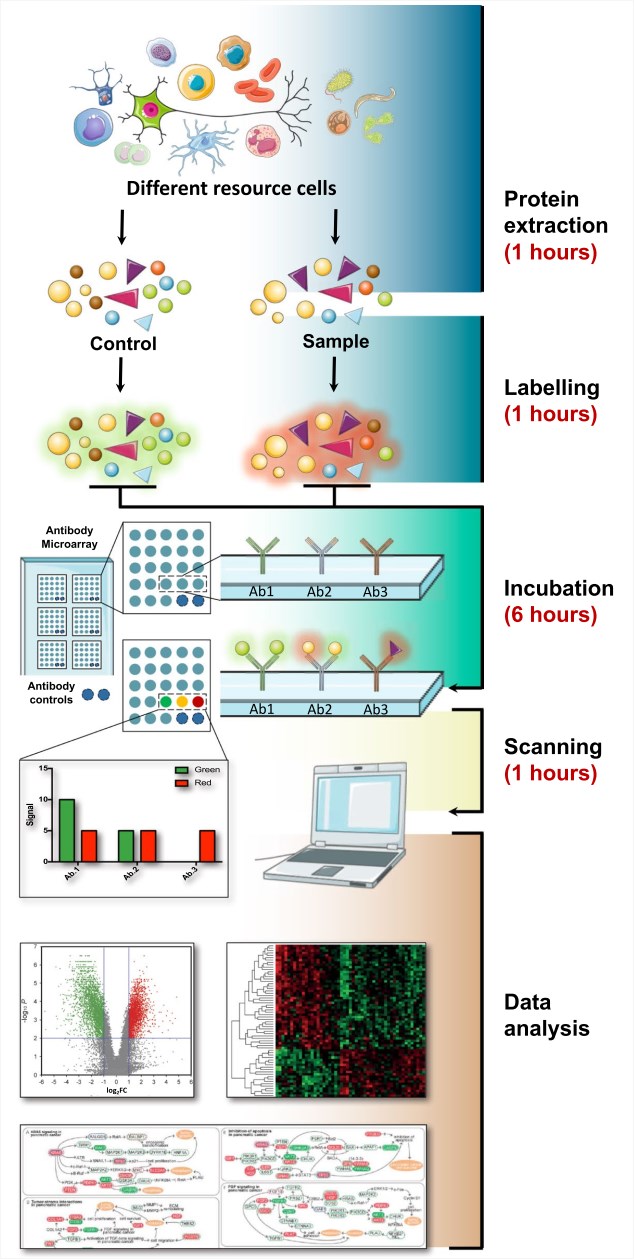 Fig.1 Review of planar antibody microarray technologies and their applications in the field of proteomics.1
Applications of Antibody Array
- Signalling Pathway Related Research
- Drug Mechanism Research
Change of signaling pathways represents a hallmark of many disease conditions, including cancer, diabetes and neurodegenerative disorders. Using antibody arrays has helped researchers to study changes in protein profiles and modifications in signaling pathways in normal biological processes and disease states through capture reagents targeting proteins in the signaling cascade of interest. A range of biological sample materials can be used in antibody array, including cell lysates, tissue extracts, and plasma.
Antibody array assays are also used to study drug mechanisms in a systematic and efficient manner. Among many possible applications, tumor-induced angiogenesis has been studied since it plays a critical role in cancer progression. For example, P11 as a novel peptide ligand with high affinity to integrin αvβ3 has been identified from a hexapeptide library (PS-SPCL) using an antibody array. The pharmacological mechanism of P11 was confirmed using a specifically designed antibody array assay containing 48 cancer-related antibodies. Results showed that P11 inhibits bFGF-induced human umbilical vein endothelial cell proliferation via mitogen-activated protein kinase and extracellular-signal regulated kinase inhibition. Additionally, the antibody array also helps to reveal that P11 can cause the upregulation of apoptotic marker p53, leading to apoptosis induction.
Clinical Autoimmune Diseases Research
Antibody arrays have been used to investigate clinical diseases, primarily analyzing serum and plasma samples. One example, the diagnosis of systemic lupus erythematosus (SLE) is difficult since its heterogeneous clinical presentation and the lack of robust biomarkers to distinguish it from active, inactive disease and other autoimmune diseases. A DotScan™ antibody microarray has been used for the screening of peripheral blood mononuclear cells from SLE patients with different disease activity, rheumatoid arthritis patients, other autoimmune disease samples, and healthy controls. The antibody array profiles enable distinguish active SLE patients from healthy controls. Using a leukocyte capture array enhanced the discriminative ability of conventional SLE diagnostics by verifying serum anti-dsDNA, complements C3 and C4, The microarrays increased the capability of discrimination of semi-active and active SLE, which will assist better disease management.
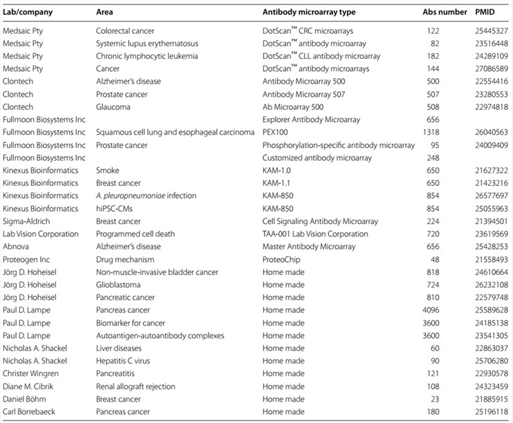 Fig.2 Review of planar antibody microarrays from recent studies.1
- Clinical Infectious Diseases Research
- Clinical Cancer Research
Antibody arrays can be used to investigate the infection mechanisms based on a proteome-wide level. For example, Helicobacter pylori is a pathogen that can infect about one half of the world’s population and result in chronic gastritis. Researcher used a DotScan™ antibody microarray to determine the tolerance of the immune system toward tumor cells in gastric cancer using 144 CD antibodies to profile the distribution of CD markers between He. pylori infected and un-infected gastric adenocarcinoma cells. Results suggested that gastric adenocarcinoma cell line AGS infected by cagA + H. pylori can increase CD27 expression, and increased CD markers were also detected in H. pylori-infected gastric cancer patients.
Furthermore, antibody arrays also are used to track a continuing change in the physiological environment, including disease status and response to therapies and interventions. Generally, after liver transplantation, about 94% of patients can be infected by hepatitis C virus (HCV) and caused histological damage even severe disease recurrence. Thus, it is urgent to find predictive biomarkers for the detection of recurrent disease severity. A CD antibody array can be used to predict the severity of HCV recurrence after transplantation. To date, five CD markers (CD27, CD182, CD260, CD41, and CD34) have been revealed that they are significantly increased in severe recurrence compared to mild recurrence. Thus, antibody arrays present a useful tool used in assessing recurrent HCV disease severity after liver transplantation.
Antibody arrays have been performed for cancer research mainly to investigate cancer progression and candidate proteins that may serve as diagnostic biomarkers. Pancreatic cancer is an aggressive disease with poor prognosis, and disease-specific biomarkers that endow early and accurate diagnosis are in urgent need. Researchers applied a recombinant antibody microarray platform have screened sera from 148 patients with pancreatic cancer, chronic pancreatitis, autoimmune pancreatitis (AIP), and healthy controls, and they have found 25 protein targets, such as IL-2, IL-11, IL-12, TNF-, which resulted in distinguishing pancreatic cancer condition from healthy controls. These targets present a high diagnostic potential. In addition, antibody arrays also are used in other cancer disease detections including lymphoma, prostate cancer, and breast cancer.
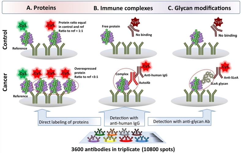 Fig.3 Antibody microarray methods to profile proteins, autoantibody-antigen complexes and glycan modifications.2
- Clinical Neurodegenerative Diseases Research
Studies for neurodegenerative pathophysiology often use the protein expression analysis. Recent advances in proteomics using antibody microarrays have offered the potential to search for novel biomarkers. Within neuroproteomics, several studies have been completed by using antibodies together with the suspension bead array assays. Profiling cerebrospinal fluid (CSF) from patients with Multiple Sclerosis (MS), researchers found GAP43 (a cytoplasmic protein) involved in the formation, and regeneration of neurons, hence a promising biomarker diseases of the brain. Compared protein levels in CSF samples collected from different neurodegenerative disease samples, the synaptic proteins GAP43 and NRGN were found to be associated with AD patients.
Current Issues and Solutions of Antibody Array
Despite the rapid development of technology, some technical issues of antibody arrays still need to be overcome to ensure high-specificity and reproducibility, and to obtain high impact data and meaningful conclusions.
For antibody microarray applications and corresponding sample types, antibody verification is required. Therefore, the data of such verification work shall be provided. At present, three main challenges need to be considered, including the quality of validated antibody, the standardization of data analysis, and the storage and sharing of data.
Proteomic analysis using antibody arrays have provided valuable data to reveal the pathophysiological background of a disease. However, the number of samples investigated in each study is usually limited. These microarray results need to be further validated with an independent set of preferably larger number of samples and by other methods. Besides, samples prepared from frozen or fresh samples may have different profiles. Another challenge of antibody microarrays for further studies is the heterozygosity of specific diseases, which needs more careful study designs, in-depth knowledge about the sample itself and the disease phenotype.
Finally, during a study using antibody microarray, control should be carefully taken into consideration. Sample controls, not only internalize control for each sample, but also positive (or negative) control also are helpful to set cut-off for further analysis.
Antibody Array Service at Creative Biolabs
Currently, a variety of forms of antibody arrays have been built as analytical tools for proteomics research. These arrays have promoted the advancement of basic biology research and discoveries towards potential tools of clinical use. Creative Biolabs provides world-class packaging antibody array service for your exact applications. Our state-of-the-art facilities and highly experienced staff are available in order to assist in arrays design and construction, as well as the data analysis of the results in a quick turnaround. If you have additional requirements or questions, please feel free to contact us.
References
-
Chen, Ziqing, et al. "Current applications of antibody microarrays." Clinical proteomics 15 (2018): 1-15.
Distributed under Open Access license CC BY 4.0, without modification. - Rho, Jung-hyun, and Paul D. Lampe. "High-throughput analysis of plasma hybrid markers for early detection of cancers." Proteomes 2.1 (2014): 1-17.