Cell Cycle Related Phosphorylation Antibody Array
The cell cycle is a fundamental evolutionarily conserved process where a cell replicates its DNA and then divides to form two daughter cells. The cell cycle comprises the S-phase (DNA synthesis phase) and M-phase (mitotic phase). Cell cycle events are controlled tightly by several kinase families including CDKs, Plks, and Aurora kinases, which directly phosphorylate and regulate hundreds of substrate proteins, and probably indirectly regulate hundreds more. These kinases collaborate, making the regulatory network much more complex but also more accurate.
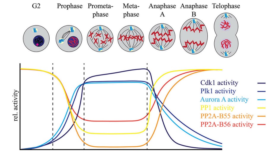 Fig.1 Fine tuning between the mitotic protein kinases and protein phosphatases regulates mitotic progression.1
Protein phosphorylation is widely recognized as the major mechanism that controls cell cycle progression, which changes downstream protein-protein interaction in such way that a signaling pathway is either switched on or shut off. Creative Biolabs offers cell cycle-related phosphorylation screening service with antibody array, which consists of 238 high-specific antibodies and more than 110 phosphorylation sites involved in cell cycle-related pathways can be tested.
Related Signaling Pathway
| Signal Pathways | |
|
|
Antibody Array Targets
| Targets |
| 14-3-3 theta/tau (S232) | CDK1 (T14) | GSK3B (S9) | p53 (S46) |
| 14-3-3 zeta (S58) | CDK2 (T160) | GSK3A/B (Y216/Y279) | p53 (S6) |
| 14-3-3 zeta/delta (T232) | CDK7 (T170) | HDAC1 (S421) | p53 (S9) |
| ABL1 (T754/T735) | Chk1 (S280) | HDAC2 (S394) | p53 (T18) |
| Abl1 (Y204) | Chk1 (S286) | HDAC3 (S424) | p53 (T81) |
| Abl1 (Y412) | Chk1 (S296) | HDAC4 (S632) | P90RSK (S380) |
| AKT (S473) | Chk1 (S301) | HDAC5 (S259) | P90RSK (T359/S363) |
| AKT (T308) | Chk1 (S317) | HDAC5 (S498) | P90RSK (T573) |
| AKT (Y326) | Chk1 (S345) | HDAC6 (S22) | p95/NBS1 (S343) |
| AKT1 (S124) | Chk2 (S516) | HDAC8 (S39) | PP2A-a (Y307) |
| AKT1 (S246) | Chk2 (T383) | Histone H2A.X (S139) | RAD51 (Y315) |
| AKT1 (T450) | Chk2 (T387) | MDM2 (S166) | RAD52 (Y104) |
| AKT1 (T72) | Chk2 (T68) | MDM4 (S367) | Rb (S608) |
| AKT1 (Y474) | CCNB1 (S126) | Myc (S373) | Rb (S780) |
| AKT2 (S474) | CCNB1 (S147) | Myc (S62) | Rb (S795) |
| ATRIP (S68/S72) | CCND1 (T286) | Myc (T358) | Rb (S807) |
| BRCA1 (S1423) | CCND3 (T283) | Myc (T58) | Rb (S811) |
| BRCA1 (S1457) | CCNE1 (T395) | p21Cip1 (T145) | Rb (T821) |
| BRCA1 (S1524) | CCNE1 (T77) | p27Kip1 (S10) | Smad2/3 (T8) |
| c-Abl (Y245) | DNA-PK (T2638) | p27Kip1 (T187) | Smad3 (S204) |
| c-Abl (Y412) | DNA-PK (T2647) | p53 (S15) | Smad3 (S208) |
| CDC2 (Y15) | E2F1 (T433) | p53 (S20) | Smad3 (S213) |
| CDC25A (S124) | FKHR (S256) | p53 (S315) | Smad3 (S425) |
| CDC25A (S75) | FKHR (S319) | p53 (S33) | Smad3 (T179) |
| CDC25B (S323) | FKHRL1 (S253) | p53 (S366) | SMC1 (S957) |
| CDC25B (S353) | FOXO1/3/4 pan (T24/T32) | p53 (S37) | TOP2A (S1106) |
| CDC25C (S216) | FOXO1A (S329) | p53 (S378) | WEE1 (S642) |
| CDC25C (T48) | FOXO1A/3A (S322/S325) | p53 (S392) |
Protocol Outline
- Extracted protein and biotinylated
- Blocked antibody array
- Incubate with treated protein and control
- Incubate with streptavidin-conjugated fluorophore
- Scan with a microarray laser scanner
- Perform densitometry and analysis
Our Analysis Service
- Suitable for protein, cell and tissue samples from human, mouse and rat
- 6×10⁷ cells or 150 µg protein
- Set replicate spots: treated sample and control
- Results are delivered in 4-5 weeks
For more detailed information about our antibody arrays, please feel free to contact us.
Reference
-
Nasa, Isha, and Arminja N. Kettenbach. "Coordination of protein kinase and phosphoprotein phosphatase activities in mitosis." Frontiers in cell and developmental biology 6 (2018): 30.
Distributed under Open Access license CC BY 4.0, without modification.
