Enzyme Linked Immunospot (ELISpot)
Overview
Enzyme-linked immunospot (ELISpot) is a technique that was developed for the detection of secreted proteins such as growth factors and cytokines. ELISpot is performed using a polyvinylidene difluoride (PVDF) or nitrocellulose (NC) membrane 96-well plate pre-coated with an antibody specific to the secreted protein. Cells are added to the plate and attach to the coated membrane. Then, cells are stimulated and the secreted protein binds to the antibody. Finally, a detection antibody is added. The resulting antibody complex can be detected either through enzymatic action to produce a colored substrate or with fluorescent tags. The membrane can be analyzed by manually counting the spots or with an automated reader designed for this purpose. Each secreting cell appears as a spot of color or fluorescence, thus this is a quantitative method for evaluating protein secretion.
As shown in Fig.1, a complete ELISpot experiment consists of three stages. It includes the isolation and preparation of the cell material to be tested, the actual assay, and the evaluation of the ELISpot plates. For the complete and detailed experimental procedure, please refer to ELISpot Protocol. We also listed some common problems and solutions in ELISpot Troubleshooting.
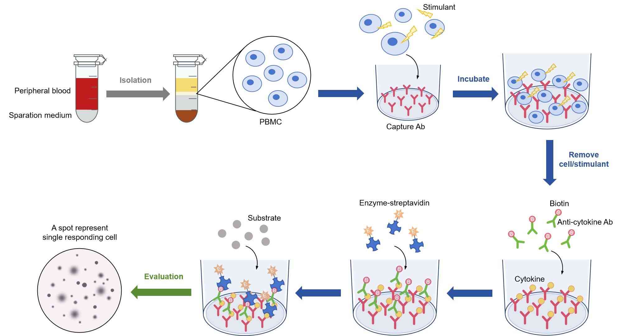 Fig.1 Three stages of an ELISpot experiment.
Conventional ELISA v.s. ELISpot
The ELISpot assay is a hybrid assay that combines the features of a mixed lymphocyte reaction (MLR) assay and an enzyme-linked immunosorbent assay (ELISA). In it's main principle, the ELISpot resembles the ELISA. In both techniques, capture antibody and detection antibody are used. However, there are more differences than similarities. An ELISA determines the total concentration of the secreted signaling protein or antibody, whereas an ELISpot detects individual cytokine or antibody secreting cells. Therefore, ELISpot should be used not 'instead of ' but rather 'in addition to' ELISA. An ELISpot assay can 100 to 400 times more sensitive than a conventional ELISA because the secreted protein is captured directly onto the well of an ELISPOT plate before having the chance to be diluted in the culture supernatant, degraded by proteases or captured by receptors on adjacent cells.
| Comparison | Conventional ELISA | ELISpot |
| Sample Format | Supernatant | Cell Culture |
| Measurement Format | Supernatant | Single Cell |
| T cell Functional Study | No | Yes |
| Per Cell Cytokine Productivity | Net Cytokine (Source Non-distinguishable) | Cytokine Per Cell (Source of Cytokine) |
| Resolution | Subtle Differences Non-detectable | High |
| Dual Detection | No | Yes |
| Storage Time | Hours | Weeks to Months |
| Detection Device | ELISA Reader | ELISpot Reader/Scanner |
Applications
The high sensitivity of ELISpot made it a powerful technique in detecting low frequency cytokine-secreting cells (about 1 in 300,000 cells) and is widely used in infections, cancer, allergies and autoimmune diseases research, vaccine development and monitor. ELISpot can be used to understand the biological effects involved in:
- Transplantation
- Vaccine development
- Th1/Th2, T cell regulation
- Monocyte and dendritic cell analysis
- Autoimmune disease
- Cancer
- Allergies
- Viral infection monitoring and treatment
Technical Tips
- We recommend the use of serum-free media for freezing, washing, and testing peripheral blood mononuclear cells (PBMCs). Even brief exposure to a mitogenic serum can cause high background while other sera can have suppressive effects.
- Avoid damage to the PVDF membrane in the wells, avoid touching the membrane with pipette tips or with the plate washer. Avoid direct contact between the well bottom and wet surfaces, including paper towels or any other materials that can absorb liquid.
- While processing plates, the PVDF membrane at the bottom of the wells must remain wet.
- Spots may not be readily visible while the membrane is still wet. Scan and count plates only after membranes have completely dried.
General Considerations for ELISpot Plate Evaluation
- Spot color and intensity of staining
- Spot gradient (staining intensity across the diameter of a spot)
- Spot size distribution
- Spot shape and definition
- Membrane coloring (= background staining of wells)
- Discrimination of spots related to their distribution and crowdedness across the well
- Occurrence of small background spot
Control Setting
- Control for background reactivity (cells alone)
- A control for false positive spots (medium alone)
- A control for overall assay functionality (cells plus mitogen)
- An internal sample control (cells plus control peptide pool, if applicable)
- An external trending control, typically consisting of 1–2 reference samples tested against medium alone, a control antigen (peptide pool against which those samples exhibit a detectable response) and mitogen.