CCN3 Antibodies
Background
CCN3 is a secreted extracellular matrix protein belonging to the CCN family, and is widely expressed in various tissues and organs. This protein plays a crucial role in tissue development, injury repair, and homeostasis maintenance by regulating processes such as cell proliferation, differentiation, and adhesion. Particularly in the heart and kidney systems, bone formation, and neural development, CCN3 participates in the regulation of organ structure and function by mediating multiple signaling pathways. Since its discovery in the early 1990s, CCN3 has received continuous attention due to its bidirectional regulatory properties in pathological processes such as fibrosis diseases, tumor occurrence, and metabolic disorders. Its multi-domain characteristic enables CCN3 to interact with various cell surface receptors and extracellular proteins, and this complex mechanism is driving the research progress in tissue regeneration medicine and disease intervention fields.
Structure of CCN3
CCN3 is a secreted extracellular matrix protein with a molecular weight of approximately 36-38 kDa. The molecular weight may vary slightly depending on different splicing variants and post-translational modifications (such as glycosylation). The following are the basic characteristics of this protein in different species:
| Species | Human | Mouse | Rat | Bovine |
|---|---|---|---|---|
| Molecular Weight (kDa) | ~38 | ~36 | ~37 | ~38 |
| Primary Structural Differences | Contains four conserved functional domains | Highly homologous domain | High similarity to the human sequence | There are species-specific modifications |
The CCN3 protein is composed of 381 amino acids (taking the human source as an example), and its primary structure contains four characteristic functional modules: the insulin-like growth factor binding protein domain, the C-type repeat sequence of von Willebrand factor, the type 1 repeat sequence of platelet reaction protein, and the cysteine-rich carboxyl-terminal domain. These modules are connected by flexible hinge regions, forming its unique multi-domain structure. The protein's secondary structure is rich in β-sheet and contains several α-helices, jointly forming its stable globular conformation. Its core function relies on the specific interactions of each domain with cell surface receptors (such as integrins) and extracellular matrix molecules, thereby regulating downstream signaling pathways.
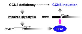 Fig. 1 Mechanism of the negative regulation of CCN3 by CCN2 in chondrocytes.1
Key structural properties of CCN3:
- Modular multi-domain structure
- Stable configuration rich in disulfide bonds
- Binding interface to multiple ligands
- Variable post-editing modifications
Functions of CCN3
CCN3 is a multifunctional signaling regulator protein. It primarily acts as a bridging molecule between the extracellular matrix and cell surface receptors, but its functions go far beyond this. The table below summarizes its main biological functions:
| Function | Description |
| Cell Proliferation Regulation | Based on the type of cells and the microenvironment, it can regulate cell proliferation in both directions. It inhibits growth in some tissues and promotes regeneration during injury repair. |
| Tissue Development and Differentiation | During the formation of bones, the development of the heart, and the establishment of the nervous system, cell differentiation and morphogenesis are coordinated through receptors such as integrins. |
| Cell Adhesion and Migration | By interacting with integrins and extracellular matrix components through its various domains, it dynamically regulates the processes of cell adhesion, spreading, and migration. |
| Vascularization Regulation | It plays a complex role in angiogenesis, being able to promote endothelial cell activity and also inhibit pathological vascular formation under certain conditions. |
| Fibrosis and Repair | It participates in the repair response after tissue damage, and its dysregulation is closely related to fibrotic lesions in organs such as the heart, kidneys, and liver. |
Unlike myoglobin, which has a clearly defined and single oxygen-binding function, the function of CCN3 is highly "context-dependent". Its biological effects depend on specific cell types, the combination of receptors bound, and local microenvironmental signals, which makes it play a complex and precise regulatory role in maintaining tissue homeostasis and responding to pathological stimuli.
Applications of CCN3 and CCN3 Antibody in Literature
1. Kubota, Satoshi, et al. "Molecular and genetic interactions between CCN2 and CCN3 behind their Yin–Yang collaboration." International journal of molecular sciences 23.11 (2022): 5887. https://doi.org/10.3390/ijms23115887
The article indicates that CCN2 and CCN3 have similar structures but antagonistic functions. In the tissue microenvironment, they interfere with each other through direct interaction and shared cofactors, forming a synergistic regulatory relationship. The interaction between the two is closely related to the regulation of sugar metabolism genes and jointly participates in processes such as cartilage development/regeneration and fibrosis.
2. Son, Seogho, et al. "CCN3/NOV promotes metastasis and tumor progression via GPNMB-induced EGFR activation in triple-negative breast cancer." Cell Death & Disease 14.2 (2023): 81. https://doi.org/10.1038/s41419-023-05608-3
The study found that in triple-negative breast cancer, CCN3 is overexpressed, which activates the Wnt/MITF axis to upregulate GPNMB, thereby promoting the activity of the EGFR/MAPK pathway, and driving the formation, growth and metastasis of tumor stem cells. CCN3 is a potential therapeutic target.
3. Xu, Nathan, Kyle Yang, and Mengjie Wang. "CCN3: lactational bone booster." Cell & Bioscience 14.1 (2024): 155. https://doi.org/10.1186/s13578-024-01344-z
Studies have shown that the CCN3 protein secreted specifically by ARH/Kiss1 neurons during lactation is an osteogenic hormone. It can activate bone stem cells and promote bone formation and repair, and is crucial for maintaining the maternal bone homeostasis and the survival of offspring.
4. Huang, Xiaojian, et al. "NOV/CCN3 induces cartilage protection by inhibiting PI3K/AKT/mTOR pathway." Journal of Cellular and Molecular Medicine 23.11 (2019): 7525-7534. https://doi.org/10.1111/jcmm.14621
The study found that the expression of CCN3 was downregulated in osteoarthritis. Supplementing with CCN3 can reduce the degradation of cartilage matrix induced by inflammatory factors by inhibiting HMGB1, activating autophagy and blocking the PI3K/AKT/mTOR pathway, indicating that it can serve as a potential therapeutic target.
5. Peng, Linan, et al. "The emerging roles of CCN3 protein in immune‐related diseases." Mediators of Inflammation 2021.1 (2021): 5576059. https://doi.org/10.1155/2021/5576059
Research has shown that CCN3 is a member of the extracellular matrix protein CCN family and regulates cell growth, migration and differentiation. Recent studies have revealed that CCN3 plays a crucial role in immune-related diseases such as rheumatoid arthritis, osteoarthritis and systemic sclerosis.
Creative Biolabs: CCN3 Antibodies for Research
Creative Biolabs specializes in the production of high-quality CCN3 antibodies for research and industrial applications. Our portfolio includes monoclonal antibodies tailored for ELISA, Flow Cytometry, Western blot, immunohistochemistry, and other diagnostic methodologies.
- Custom CCN3 Antibody Development: Tailor-made solutions to meet specific research requirements.
- Bulk Production: Large-scale antibody manufacturing for industry partners.
- Technical Support: Expert consultation for protocol optimization and troubleshooting.
- Aliquoting Services: Conveniently sized aliquots for long-term storage and consistent experimental outcomes.
For more details on our CCN3 antibodies, custom preparations, or technical support, contact us at email.
Reference
- Kubota, Satoshi, et al. "Molecular and genetic interactions between CCN2 and CCN3 behind their Yin–Yang collaboration." International journal of molecular sciences 23.11 (2022): 5887. Distributed under Open Access license CC BY 4.0, without modification.https://doi.org/10.3390/ijms23115887
Anti-CCN3 antibodies
 Loading...


Hot products 
-
Mouse Anti-APOE Recombinant Antibody (A1) (CBMAB-0078CQ)

-
Mouse Anti-CCS Recombinant Antibody (CBFYC-1093) (CBMAB-C1150-FY)

-
Mouse Anti-AHCYL1 Recombinant Antibody (V2-180270) (CBMAB-A1703-YC)

-
Mouse Anti-8-oxoguanine Recombinant Antibody (V2-7697) (CBMAB-1869CQ)

-
Mouse Anti-CHRNA9 Recombinant Antibody (8E4) (CBMAB-C9161-LY)

-
Mouse Anti-FOXL1 Recombinant Antibody (CBXF-0845) (CBMAB-F0462-CQ)

-
Mouse Anti-CDKL5 Recombinant Antibody (CBFYC-1629) (CBMAB-C1689-FY)

-
Mouse Anti-ARIH1 Recombinant Antibody (C-7) (CBMAB-A3563-YC)

-
Mouse Anti-CFL1 Recombinant Antibody (CBFYC-1771) (CBMAB-C1833-FY)

-
Mouse Anti-APOA1 Monoclonal Antibody (CBFYR0637) (CBMAB-R0637-FY)

-
Human Anti-SARS-CoV-2 S1 Monoclonal Antibody (CBFYR-0120) (CBMAB-R0120-FY)

-
Mouse Anti-DHFR Recombinant Antibody (D0821) (CBMAB-D0821-YC)

-
Mouse Anti-AKT1 Recombinant Antibody (V2-180546) (CBMAB-A2070-YC)

-
Mouse Anti-AK4 Recombinant Antibody (V2-180419) (CBMAB-A1891-YC)

-
Mouse Anti-EMP3 Recombinant Antibody (CBFYE-0100) (CBMAB-E0207-FY)

-
Mouse Anti-ACE2 Recombinant Antibody (V2-179293) (CBMAB-A0566-YC)

-
Mouse Anti-F11R Recombinant Antibody (402) (CBMAB-0026-WJ)

-
Rat Anti-(1-5)-α-L-Arabinan Recombinant Antibody (V2-501861) (CBMAB-XB0003-YC)

-
Mouse Anti-CD24 Recombinant Antibody (ALB9) (CBMAB-0176CQ)

-
Mouse Anti-ACTN4 Recombinant Antibody (V2-6075) (CBMAB-0020CQ)

- AActivation
- AGAgonist
- APApoptosis
- BBlocking
- BABioassay
- BIBioimaging
- CImmunohistochemistry-Frozen Sections
- CIChromatin Immunoprecipitation
- CTCytotoxicity
- CSCostimulation
- DDepletion
- DBDot Blot
- EELISA
- ECELISA(Cap)
- EDELISA(Det)
- ESELISpot
- EMElectron Microscopy
- FFlow Cytometry
- FNFunction Assay
- GSGel Supershift
- IInhibition
- IAEnzyme Immunoassay
- ICImmunocytochemistry
- IDImmunodiffusion
- IEImmunoelectrophoresis
- IFImmunofluorescence
- IGImmunochromatography
- IHImmunohistochemistry
- IMImmunomicroscopy
- IOImmunoassay
- IPImmunoprecipitation
- ISIntracellular Staining for Flow Cytometry
- LALuminex Assay
- LFLateral Flow Immunoassay
- MMicroarray
- MCMass Cytometry/CyTOF
- MDMeDIP
- MSElectrophoretic Mobility Shift Assay
- NNeutralization
- PImmunohistologyp-Paraffin Sections
- PAPeptide Array
- PEPeptide ELISA
- PLProximity Ligation Assay
- RRadioimmunoassay
- SStimulation
- SESandwich ELISA
- SHIn situ hybridization
- TCTissue Culture
- WBWestern Blot
