CD3E Antibodies
Background
The CD3E gene encodes the CD3ε chain of the surface glycoprotein of T cells, which is an important component of the T cell receptor complex. This protein participates in the formation of the immune synapse by binding to the TCR, transmitting the antigen recognition signal to the cell interior, thereby activating the immune response function of T cells. Deficiency of the CD3E gene can lead to severe combined immunodeficiency disease, with clinical manifestations including recurrent infections and immune dysfunction. This gene was first identified in 1986, and its three-dimensional structure was resolved through cryo-electron microscopy technology in the late 2000s, providing a key basis for understanding the molecular mechanism of adaptive immunity. Antibody drugs targeting CD3E have been developed for use in organ transplantation anti-rejection and the treatment of autoimmune diseases, and the research on its molecular mechanism continues to drive the development of immunology and clinical treatment fields.
Structure of CD3E
The CD3ε protein encoded by the CD3E gene is a transmembrane glycoprotein with a molecular weight of approximately 19-26 kDa. Its precise molecular weight varies among different species, mainly due to the differences in the degree of glycosylation modification.
| Species | Human | Mouse | Rat |
|---|---|---|---|
| Molecular Weight (kDa) | 19 - 26 | 20 - 25 | 19 - 25 |
| Primary Structural Differences | Containing an extracellular domain Ig sample structure, a transmembrane region and intracellular tail, a long intracellular tail contains an immune receptor tyrosine motif (ITAM). | Highly homologous to humans, the intracellular ITAM sequence is highly conserved and is the key to signal transduction. | Domain composition is generally consistent with functional regions and human, with positively charged arginine in the transmembrane region involved and assembly of the TCR complex. |
This protein is a core component of the T-cell receptor (TCR) complex. Its extracellular part assists in the assembly and positioning of the TCR, while the immunoreceptor tyrosine activation motif (ITAM) in its intracellular region is phosphorylated after the TCR recognizes the antigen, and it is a key molecular switch that initiates the downstream signal cascade and ultimately activates T cells.
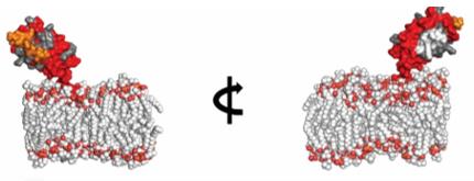 Fig. 1 Structural model of human CD3E alone in the membrane.1
Key structural properties of CD3E:
- Extracellular Ig-like single domain, responsible for binding to other TCR subunits
- The transmembrane region with positive charge
- Intracellular segment contains conserved immune receptor tyrosine activating motifs (ITAM)
- Highly conserved cysteine residues form intramolecular disulfide bonds
Functions of CD3E
The protein encoded by the CD3E gene is a key signaling subunit of the T-cell receptor (TCR) complex. Its core function is to convert the extracellular antigen recognition signal into an intracellular activation signal, thereby initiating the adaptive immune response.
| Function | Description |
| Signal transduction initiation | The ITAM motif in the intracellular region is phosphorylated after TCR binds to the antigen, recruiting and activating kinases such as ZAP-70, which serves as the starting point of the T cell activation cascade. |
| Assembly and Stability of TCR Complex | The transmembrane region of the TCR interacts with other CD3 subunits and TCR chains through electrostatic interactions, which is essential for the correct assembly and expression of the complete receptor complex on the membrane. |
| Signal Amplification and Regulation | The degree and pattern of phosphorylation of ITAM can regulate the intensity of the signal, influencing the activation threshold of T cells. It plays a crucial regulatory role in thymic selection and peripheral tolerance. |
| Immune Synapse Formation | The tissues involved in the contact interface between T cells and antigen-presenting cells (immune synapse) bring the TCR-CD3 complex to the signal center, enhancing the signal efficiency. |
| Clinical Target Application | As a characteristic surface protein of T cells, it serves as the target for anti-CD3 monoclonal antibodies (such as OKT3), and is used in clinical immunosuppressive therapy and CAR-T cell modification. |
Unlike multi-subunit signal complexes with synergistic effects, the signal initiated by the single ITAM motif of CD3ε is the initial and crucial one. The phosphorylation events of this motif determine the fate of the T cell response and serves as the core hub connecting antigen recognition and cell function execution.
Applications of CD3E and CD3E Antibody in Literature
1. Zorn, Julie A., et al. "Humanization of a strategic CD3 epitope enables evaluation of clinical T-cell engagers in a fully immunocompetent in vivo model." Scientific Reports 12.1 (2022): 3530. https://doi.org/10.1038/s41598-022-06953-7
This article introduces a new hCD3E-epi mouse model, in which T cells can be activated by human CD3E antibodies. This model enables the evaluation of the anti-tumor efficacy of human T cell connectors (TCE) in immunocompetent hosts, providing a research tool that is closer to the human immune environment for preclinical studies.
2. Liu, Yi, et al. "CXCL12 and CD3E as indicators for tumor microenvironment modulation in bladder cancer and their correlations with immune infiltration and molecular subtypes." Frontiers in oncology 11 (2021): 636870. https://doi.org/10.3389/fonc.2021.636870
By analyzing the data of bladder cancer patients, it was found that high expression of CXCL12 and low expression of CD3E were associated with a longer survival period, and both were related to the infiltration of specific immune cells in the tumor microenvironment (TME), suggesting that they could serve as potential therapeutic targets.
3. Aguilera-Braico, Diego Maximo, and Gabriela A. Balogh. "CD3Ɛ immune restorative ability induced by Maitake Pro4x in immunosupressed BALBc mice." BMC Research Notes 15.1 (2022): 307. https://doi.org/10.1186/s13104-022-06201-1
This study shows that Maitake Pro4X can significantly restore the number of CD3Ɛ T cells in the lymph nodes of BALBc mice that were inhibited by dexamethasone (with a recovery rate of 42.97%, p < 0.01), and increase the level of the Ly6G cell population, confirming its ability to reverse immune suppression.
4. Kim, Shihyoung, et al. "Comparison of CD3e antibody and CD3e-sZAP immunotoxin treatment in mice identifies sZAP as the main driver of vascular leakage." Biomedicines 10.6 (2022): 1221. https://doi.org/10.3390/biomedicines10061221
This study compared non-mitogenic CD3ε monoclonal antibody with the new CD3ε immunotoxin. The research found that the vascular leakage syndrome triggered by the immunotoxin was mainly driven by its toxin component rather than the CD3ε targeting component. This provides a direction for improving the safety of CD3ε-targeted therapies.
5. Zheng, Xiaonan, et al. "LCK and CD3E orchestrate the tumor microenvironment and promote immunotherapy response and survival of muscle-invasive bladder cancer patients." Frontiers in cell and developmental biology 9 (2021): 748280. https://doi.org/10.3389/fcell.2021.748280
This study analyzed multiple sets of data to determine that CD3E and LCK are potential biomarkers for prognosis and immunotherapy efficacy in muscle-invasive bladder cancer. Their high expression is positively correlated with better survival rates and immunotherapy response rates, and is also associated with the expression of various immune checkpoints.
Creative Biolabs: CD3E Antibodies for Research
Creative Biolabs specializes in the production of high-quality CD3E antibodies for research and industrial applications. Our portfolio includes monoclonal antibodies tailored for ELISA, Flow Cytometry, Western blot, immunohistochemistry, and other diagnostic methodologies.
- Custom CD3E Antibody Development: Tailor-made solutions to meet specific research requirements.
- Bulk Production: Large-scale antibody manufacturing for industry partners.
- Technical Support: Expert consultation for protocol optimization and troubleshooting.
- Aliquoting Services: Conveniently sized aliquots for long-term storage and consistent experimental outcomes.
For more details on our CD3E antibodies, custom preparations, or technical support, contact us at email.
Reference
- Zorn, Julie A., et al. "Humanization of a strategic CD3 epitope enables evaluation of clinical T-cell engagers in a fully immunocompetent in vivo model." Scientific Reports 12.1 (2022): 3530. Distributed under the same Creative Commons license CC BY 4.0 as the original. Cropped from the original figure.https://doi.org/10.1038/s41598-022-06953-7
Anti-CD3E antibodies
 Loading...


Hot products 
-
Mouse Anti-BCL6 Recombinant Antibody (CBYY-0442) (CBMAB-0445-YY)

-
Mouse Anti-CSPG4 Recombinant Antibody (CBFYM-1050) (CBMAB-M1203-FY)

-
Mouse Anti-CCND2 Recombinant Antibody (DCS-3) (CBMAB-G1318-LY)

-
Mouse Anti-BrdU Recombinant Antibody (IIB5) (CBMAB-1038CQ)

-
Rabbit Anti-BAD (Phospho-Ser136) Recombinant Antibody (CAP219) (CBMAB-AP536LY)

-
Mouse Anti-BZLF1 Recombinant Antibody (BZ.1) (CBMAB-AP705LY)

-
Mouse Anti-Acetyl-α-Tubulin (Lys40) Recombinant Antibody (V2-623485) (CBMAB-CP2897-LY)

-
Mouse Anti-BCL2L1 Recombinant Antibody (H5) (CBMAB-1025CQ)

-
Mouse Anti-ATP1A2 Recombinant Antibody (M7-PB-E9) (CBMAB-A4013-YC)

-
Rabbit Anti-ABL1 (Phosphorylated Y245) Recombinant Antibody (V2-505716) (PTM-CBMAB-0465LY)

-
Mouse Anti-BAX Recombinant Antibody (CBYY-0216) (CBMAB-0217-YY)

-
Mouse Anti-ANXA7 Recombinant Antibody (A-1) (CBMAB-A2941-YC)

-
Mouse Anti-ASH1L Monoclonal Antibody (ASH5H03) (CBMAB-1372-YC)

-
Mouse Anti-BIRC3 Recombinant Antibody (16E63) (CBMAB-C3367-LY)

-
Rabbit Anti-Acetyl-Histone H4 (Lys16) Recombinant Antibody (V2-623415) (CBMAB-CP1021-LY)

-
Mouse Anti-ADGRE5 Recombinant Antibody (V2-360335) (CBMAB-C2088-CQ)

-
Mouse Anti-AAV-5 Recombinant Antibody (V2-503416) (CBMAB-V208-1402-FY)

-
Mouse Anti-DES Monoclonal Antibody (440) (CBMAB-AP1857LY)

-
Mouse Anti-CALR Recombinant Antibody (CBFYC-0763) (CBMAB-C0818-FY)

-
Mouse Anti-C5AR1 Recombinant Antibody (R63) (CBMAB-C9553-LY)

- AActivation
- AGAgonist
- APApoptosis
- BBlocking
- BABioassay
- BIBioimaging
- CImmunohistochemistry-Frozen Sections
- CIChromatin Immunoprecipitation
- CTCytotoxicity
- CSCostimulation
- DDepletion
- DBDot Blot
- EELISA
- ECELISA(Cap)
- EDELISA(Det)
- ESELISpot
- EMElectron Microscopy
- FFlow Cytometry
- FNFunction Assay
- GSGel Supershift
- IInhibition
- IAEnzyme Immunoassay
- ICImmunocytochemistry
- IDImmunodiffusion
- IEImmunoelectrophoresis
- IFImmunofluorescence
- IGImmunochromatography
- IHImmunohistochemistry
- IMImmunomicroscopy
- IOImmunoassay
- IPImmunoprecipitation
- ISIntracellular Staining for Flow Cytometry
- LALuminex Assay
- LFLateral Flow Immunoassay
- MMicroarray
- MCMass Cytometry/CyTOF
- MDMeDIP
- MSElectrophoretic Mobility Shift Assay
- NNeutralization
- PImmunohistologyp-Paraffin Sections
- PAPeptide Array
- PEPeptide ELISA
- PLProximity Ligation Assay
- RRadioimmunoassay
- SStimulation
- SESandwich ELISA
- SHIn situ hybridization
- TCTissue Culture
- WBWestern Blot
