CD55 Antibodies
Background
CD55 is a complement regulatory protein widely expressed on the surface of human cell membranes and belongs to the complement activation regulatory factor family. This protein maintains immune homeostasis by inhibiting the formation and stability of complement C3/C5 convertase, protecting normal cells from accidental damage by the complement system. It has significant clinical significance in autoimmune diseases such as paroxysmal nocturnal hemoglobinuria, which causes red blood cells to be sensitive to complement due to the absence of GPI anchor proteins such as CD55. First discovered on red blood cell membranes in the 1970s, CD55 is also one of the first complement regulatory proteins to be clarified, and its crystal structure was analyzed in the early 21st century. In-depth research on the mechanism of action of CD55 has greatly promoted the development of strategies for regulating the complement system, cellular immune defense, and treating autoimmune diseases.
Structure of CD55
CD55 is a glycosylated membrane protein with a relatively small molecular weight. The molecular weight of its core polypeptide is approximately 35-40 kDa. However, due to different degrees of glycosylation, the actual molecular weight varies within the range of 55-75 kDa. This difference mainly stems from the degree of modification of multiple N-linked and O-linked glycosylation sites in its amino acid sequence.
| Species | Human | Mouse | Bovine | Pig |
| Molecular Weight (kDa) | 70-75 | 65-70 | 68-72 | 66-70 |
| Primary Structural Differences | Four SCR domains, GPI anchored | Highly homologous to humans, GPI anchored | SCR structure domain number conservative | Glycosylation pattern interspecific differences |
This protein is composed of a polypeptide chain of approximately 350 amino acid residues, and its primary structure folds into a characteristic short common repeat sequence (SCR domain), usually containing four such domains. The protein structure of CD55 is anchored to the cell membrane by a glycosylphosphatidylinositol (GPI) at the C-terminal, and this anchoring method makes it fluid on the membrane. The SCR domain at its N-terminal is responsible for interacting with complement proteins and performs regulatory functions by accelerating the decay of C3/C5 convertase. The N-linked and O-linked sugar chains on the surface of proteins not only contribute to their apparent molecular weight, but also directly participate in their specific recognition with complement components.
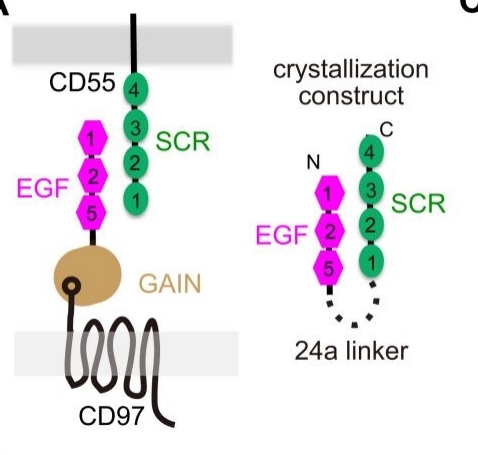 Fig. 1 The CD97–CD55 binding pattern and our chimeric construct.1
Key structural properties of CD55:
- By multiple short of tandem repeat (SCR) structure domain constitute extracellular region
- Anchored to the cell membrane surface by glycosylphosphatidylinositol (GPI)
- SCR domains mediate specific binding to complement components C3/C4b
Functions of CD55
The core function of CD55 is to protect host cells from accidental damage by the complement system, and its mechanism of action also involves multiple immune regulatory processes.
| Function | Description |
| Complement inhibition | By accelerating the decay of C3/C5 convertase or promoting its degradation, it blocks the complement activation cascade reaction and protects normal cells from damage by membrane attack complexes. |
| Maintenance of immune homeostasis | Forming a protective layer in the surface of red blood cells, platelets and endothelial cells, maintaining the complement system appears in pathogen removal and its fine balance between the organization attack. |
| Cell signal regulation | In addition to complement regulation, CD55 can also participate in processes such as cell migration, proliferation and inflammatory response by interacting with ligands like CD97. |
| Disease defense | In diseases such as paroxysmal nocturnal hemoglobinuria, the loss of CD55 expression leads to a significant increase in the complement sensitivity of red blood cells, causing intravascular hemolysis. |
| Reproductive and developmental support | It is highly expressed in the trophoblast cells of the placenta, protecting fetal tissues from being attacked by the maternal complement system and playing an important role in maintaining pregnancy. |
The regulation of complement by CD55 does not rely on other cofactors. This autonomous mechanism of action enables it to respond rapidly to complement activation signals in the microenvironment, ensuring the immediality of cell protection.
Applications of CD55 and CD55 Antibody in Literature
1. Shakya, Bikash, et al. "Erythrocyte CD55 mediates the internalization of Plasmodium falciparum parasites." Elife 10 (2021): e61516. https://doi.org/10.7554/eLife.61516
The article indicates that research on the invasion of red blood cells by Plasmodium falciparum has found that CD55 is a key factor in the internalization process of the parasite. This protein plays a role in the mobile connection stage, does not affect the previous attachment but is necessary for invasion. Its function is broad-spectrum, suggesting that CD55 and related pathways can serve as potential therapeutic targets for malaria.
2. Li, L., et al. "CD55 is over-expressed in the tumour environment." British journal of cancer 84.1 (2001): 80-86. https://doi.org/10.1054/bjoc.2000.1570
Studies have shown that CD55 is significantly upregulated in tumor cells and stroma. The CD55 on the surface of tumor cells can be a hundred times higher than that of normal cells and can be deposited in the extracellular matrix. This high expression characteristic in the tumor environment makes it an ideal target for tumor imaging and immunotherapy.
3. Mikesch, Jan-Henrik, et al. "The expression and action of decay‐accelerating factor (CD55) in human malignancies and cancer therapy." Analytical Cellular Pathology 28.5-6 (2006): 223-232. https://doi.org/10.1155/2006/814816
Studies have shown that CD55 is highly expressed in various malignant tumors, and its function far exceeds immune regulation. It promotes tumorigenesis, angiogenesis, metastasis and helps tumor cells escape through multiple signaling pathways, and thus has become a highly promising new target for cancer treatment.
4. Gaykema, Lonneke H., et al. "Inhibition of complement activation by CD55 overexpression in human induced pluripotent stem cell derived kidney organoids." Frontiers in Immunology 13 (2023): 1058763. https://doi.org/10.3389/fimmu.2022.1058763
Studies have shown that to address immune rejection in stem cell-derived kidney transplantation, human-induced pluripotent stem cell lines overexpressing CD55 have been constructed. The results show that CD55 can effectively inhibit complement activation, providing a novel protective strategy against humoral immunity for tissue transplantation.
5. Kaneko, Kazuyo, et al. "The active form of Helicobacter pylori vacuolating cytotoxin induces decay‐accelerating factor CD55 in association with intestinal metaplasia in the human gastric mucosa." The Journal of pathology 258.2 (2022): 199-209. https://doi.org/10.1002/path.5990
Studies have shown that infection with Helicobacter pylori vacA i1 strain can induce an increase in the expression of CD55 in gastric mucosa and is associated with intestinal metaplasia. CD55 partially regulates inflammatory responses in gastric epithelial cells, suggesting that it may become a potential target in intestinal metaplasia and gastric cancer management.
Creative Biolabs: CD55 Antibodies for Research
Creative Biolabs specializes in the production of high-quality CD55 antibodies for research and industrial applications. Our portfolio includes monoclonal antibodies tailored for ELISA, Flow Cytometry, Western blot, immunohistochemistry, and other diagnostic methodologies.
- Custom CD55 Antibody Development: Tailor-made solutions to meet specific research requirements.
- Bulk Production: Large-scale antibody manufacturing for industry partners.
- Technical Support: Expert consultation for protocol optimization and troubleshooting.
- Aliquoting Services: Conveniently sized aliquots for long-term storage and consistent experimental outcomes.
For more details on our CD55 antibodies, custom preparations, or technical support, contact us at email.
Reference
- Niu, Minghui, et al. "Structural basis for CD97 recognition of the decay-accelerating factor CD55 suggests mechanosensitive activation of adhesion GPCRs." Journal of Biological Chemistry 296 (2021). https://doi.org/10.1016/j.jbc.2021.100776
Anti-CD55 antibodies
 Loading...


Hot products 
-
Mouse Anti-BMI1 Recombinant Antibody (CBYC-P026) (CBMAB-P0108-YC)

-
Mouse Anti-C5AR1 Recombinant Antibody (R63) (CBMAB-C9553-LY)

-
Mouse Anti-FOSB Recombinant Antibody (CBXF-3593) (CBMAB-F2522-CQ)

-
Rat Anti-ADGRE4 Recombinant Antibody (V2-160163) (CBMAB-F0011-CQ)

-
Mouse Anti-FN1 Monoclonal Antibody (71) (CBMAB-1241CQ)

-
Mouse Anti-CALR Recombinant Antibody (CBFYC-0763) (CBMAB-C0818-FY)

-
Mouse Anti-HTLV-1 gp46 Recombinant Antibody (CBMW-H1006) (CBMAB-V208-1154-FY)

-
Mouse Anti-GFAP Recombinant Antibody (5) (CBMAB-G0346-LY)

-
Rat Anti-CD63 Recombinant Antibody (7G4.2E8) (CBMAB-C8725-LY)

-
Mouse Anti-ATP1B3 Recombinant Antibody (1E9) (CBMAB-A4021-YC)

-
Rabbit Anti-CCN1 Recombinant Antibody (CBWJC-3580) (CBMAB-C4816WJ)

-
Mouse Anti-ACE2 Recombinant Antibody (V2-179293) (CBMAB-A0566-YC)

-
Mouse Anti-FTH1 Recombinant Antibody (CBXF-1896) (CBMAB-F3426-CQ)

-
Mouse Anti-APOA1 Monoclonal Antibody (CBFYR0637) (CBMAB-R0637-FY)

-
Mouse Anti-ACVR1C Recombinant Antibody (V2-179685) (CBMAB-A1041-YC)

-
Mouse Anti-ADGRL2 Recombinant Antibody (V2-58519) (CBMAB-L0166-YJ)

-
Mouse Anti-ARIH1 Recombinant Antibody (C-7) (CBMAB-A3563-YC)

-
Mouse Anti-CRYAB Recombinant Antibody (A4345) (CBMAB-A4345-YC)

-
Mouse Anti-4-Hydroxynonenal Recombinant Antibody (V2-502280) (CBMAB-C1055-CN)

-
Rabbit Anti-ALDOA Recombinant Antibody (D73H4) (CBMAB-A2314-YC)

- AActivation
- AGAgonist
- APApoptosis
- BBlocking
- BABioassay
- BIBioimaging
- CImmunohistochemistry-Frozen Sections
- CIChromatin Immunoprecipitation
- CTCytotoxicity
- CSCostimulation
- DDepletion
- DBDot Blot
- EELISA
- ECELISA(Cap)
- EDELISA(Det)
- ESELISpot
- EMElectron Microscopy
- FFlow Cytometry
- FNFunction Assay
- GSGel Supershift
- IInhibition
- IAEnzyme Immunoassay
- ICImmunocytochemistry
- IDImmunodiffusion
- IEImmunoelectrophoresis
- IFImmunofluorescence
- IGImmunochromatography
- IHImmunohistochemistry
- IMImmunomicroscopy
- IOImmunoassay
- IPImmunoprecipitation
- ISIntracellular Staining for Flow Cytometry
- LALuminex Assay
- LFLateral Flow Immunoassay
- MMicroarray
- MCMass Cytometry/CyTOF
- MDMeDIP
- MSElectrophoretic Mobility Shift Assay
- NNeutralization
- PImmunohistologyp-Paraffin Sections
- PAPeptide Array
- PEPeptide ELISA
- PLProximity Ligation Assay
- RRadioimmunoassay
- SStimulation
- SESandwich ELISA
- SHIn situ hybridization
- TCTissue Culture
- WBWestern Blot
