S100B Antibodies
Background
The S100B gene encodes a calcium-binding protein that is mainly expressed specifically by astrocytes in the central nervous system. This protein performs multiple biological functions in the form of a homodimer. It maintains the normal physiological activities of the nervous system by regulating intracellular calcium homeostasis and participating in cell proliferation and differentiation processes. At the same time, it is released into cerebrospinal fluid and peripheral blood under pathological conditions such as brain injury and neurodegenerative diseases. This gene was discovered in 1965 and is named for its 100% solubility in saturated ammonium sulfate solution. It is one of the members of the S100 protein family that has been deeply studied. Its stable dimer structure and specific expression pattern have become important biomarkers for the diagnosis of nerve injury and provide key molecular models for the study of neuronutrition, apoptosis mechanisms and glial cell functions.
Structure of S100B
S100B is a calcium-binding protein with a molecular weight of approximately 10.4 kDa. Its precise molecular weight varies slightly among different species due to differences in amino acid composition.
| Species | Human | Mouse | Rat |
| Molecular Weight (kDa) | 10.4 | 10.5 | 10.4 |
| Primary Structural Differences | Conservative EF hand calcium-binding domain | Highly homologous to humans | One key glutamine site difference |
This protein is a dimer formed by two identical subunits through cysteine, and its three-dimensional structure contains four α -helices and two EF hand structure mods. Each subunit can bind to two calcium ions through a hydrophobic pocket, and the conformational change exposes the target binding site. The C-terminal extension region contains specific residues that interact with p53, and disulfide bonds play a decisive role in maintaining the stability of the protein's quaternary structure.
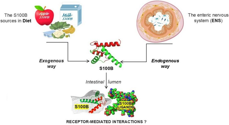 Fig. 1 Schematic diagram of the endogenous and exogenous sources of S100B.1
Key structural properties of S100B:
- Stable homodimer configuration
- Each subunit contains two EF hand calcium-binding motifs
- Conformational changes induced by calcium ions expose hydrophobic binding sites
Functions of S100B
The core function of the S100B protein is to maintain intracellular calcium homeostasis and participate in signal transduction, while it exhibits dual biological activity at different concentrations.
| Function | Description |
| Calcium signal regulation | By binding to calcium ions, conformational changes occur, thereby regulating cell proliferation, differentiation and metabolic processes. |
| Neurotrophic effect | The molar concentration of promote the growth of neurons and synaptic plasticity, is conducive to the development of the nervous system to repair. |
| Neuroinflammatory promotion | At micro-molar concentrations, it can activate pathways such as NF-κB, intensify the inflammatory response of glial cells, and participate in neuropathological processes. |
| Regulation of apoptosis | By p53 dependent and not rely on the way to affect the process of cell cycle and apoptosis, the role has the concentration dependence. |
| Biomarker function | In brain damage, release to peripheral blood in neurodegenerative diseases, has become an important molecular diagnosis index. |
The biological effects of S100B show a significant concentration dependence: at low concentrations, it exerts neurotrophic and protective effects, while at high concentrations, it exhibits pro-inflammatory and neurotoxic properties. This dual characteristic is known as the "S100B paradox".
Applications of S100B and S100B Antibody in Literature
1. Oris, Charlotte, et al. "S100B, actor and biomarker of mild traumatic brain injury." International Journal of Molecular Sciences 24.7 (2023): 6602. https://doi.org/10.3390/ijms24076602
The article indicates that S100B is a key biomarker in the diagnosis and treatment of mild brain injury. Its detection in the acute phase can help reduce unnecessary imaging examinations and hospitalizations, and may become a potential therapeutic target.
2. Zheng, Zhen, Peng Zheng, and Xiaobing Zou. "Peripheral blood S100B levels in autism spectrum disorder: a systematic review and meta-analysis." Journal of Autism and Developmental Disorders 51.8 (2021): 2569-2577. https://doi.org/10.1007/s10803-020-04710-1
This study, through meta-analysis, found that the level of S100B protein in the peripheral blood of patients with autism spectrum disorder (ASD) was significantly higher than that of healthy people, indicating that S100B has the potential to become a biomarker for the auxiliary diagnosis of ASD.
3. Michetti, Fabrizio, and Vincenzo Romano Spica. "The "Jekyll Side" of the S100B Protein: Its Trophic Action in the Diet." Nutrients 17.5 (2025): 881. https://doi.org/10.3390/nu17050881
The article indicates that the function of S100B protein has a dual nature of concentration: at low concentrations, it exerts beneficial neurotrophic effects, while at high concentrations, it produces pro-inflammatory/toxic effects. The latest research has found that it exists as a nutrient in breast milk and functions by influencing the microbiota, providing a new direction for the study of its functions.
4. Lai, Pui Man Rosalind, and Rose Du. "Association between S100B levels and long-term outcome after aneurysmal subarachnoid hemorrhage: systematic review and pooled analysis." PLoS One 11.3 (2016): e0151853. https://doi.org/10.1371/journal.pone.0151853
Studies have shown that elevated serum S100B protein levels are significantly associated with the risk of cerebral infarction and poor prognosis in patients with aneurysmal subarachnoid hemorrhage, suggesting that S100B can serve as a potential biomarker for evaluating the prognosis of this disease.
5. Teurneau-Hermansson, Karl, et al. "S100B predicts neurological injury and 30-day mortality following surgery for acute type A aortic dissection: an observational cohort study." Journal of Cardiothoracic Surgery 18.1 (2023): 62. https://doi.org/10.1186/s13019-023-02151-2
Research has found that the serum S100B level 24 hours after surgery is A strong independent predictor of nerve injury and 30-day mortality after type A aortic dissection surgery. The risk significantly increases when the level is ≥0.23μg/l.
Creative Biolabs: S100B Antibodies for Research
Creative Biolabs specializes in the production of high-quality S100B antibodies for research and industrial applications. Our portfolio includes monoclonal antibodies tailored for ELISA, Flow Cytometry, Western blot, immunohistochemistry, and other diagnostic methodologies.
- Custom S100B Antibody Development: Tailor-made solutions to meet specific research requirements.
- Bulk Production: Large-scale antibody manufacturing for industry partners.
- Technical Support: Expert consultation for protocol optimization and troubleshooting.
- Aliquoting Services: Conveniently sized aliquots for long-term storage and consistent experimental outcomes.
For more details on our S100B antibodies, custom preparations, or technical support, contact us at email.
Reference
- Michetti, Fabrizio, and Vincenzo Romano Spica. "The "Jekyll Side" of the S100B Protein: Its Trophic Action in the Diet." Nutrients 17.5 (2025): 881. https://doi.org/10.3390/nu17050881
Anti-S100B antibodies
 Loading...


Hot products 
-
Mouse Anti-DMD Recombinant Antibody (D1190) (CBMAB-D1190-YC)

-
Mouse Anti-CCT6A/B Recombinant Antibody (CBXC-0168) (CBMAB-C5570-CQ)

-
Mouse Anti-CECR2 Recombinant Antibody (CBWJC-2465) (CBMAB-C3533WJ)

-
Mouse Anti-2C TCR Recombinant Antibody (V2-1556) (CBMAB-0951-LY)

-
Mouse Anti-14-3-3 Pan Recombinant Antibody (V2-9272) (CBMAB-1181-LY)

-
Mouse Anti-BSN Recombinant Antibody (219E1) (CBMAB-1228-CN)

-
Mouse Anti-CD83 Recombinant Antibody (HB15) (CBMAB-C1765-CQ)

-
Mouse Anti-ATG5 Recombinant Antibody (9H197) (CBMAB-A3945-YC)

-
Mouse Anti-CASQ1 Recombinant Antibody (CBFYC-0863) (CBMAB-C0918-FY)

-
Mouse Anti-ACO2 Recombinant Antibody (V2-179329) (CBMAB-A0627-YC)

-
Mouse Anti-AMACR Recombinant Antibody (CB34A) (CBMAB-CA034LY)

-
Mouse Anti-DISP2 Monoclonal Antibody (F66A4B1) (CBMAB-1112CQ)

-
Rat Anti-ABCC11 Recombinant Antibody (V2-179001) (CBMAB-A0236-YC)

-
Rabbit Anti-CCN1 Recombinant Antibody (CBWJC-3580) (CBMAB-C4816WJ)

-
Mouse Anti-APCS Recombinant Antibody (CBYC-A663) (CBMAB-A3054-YC)

-
Rabbit Anti-DLK1 Recombinant Antibody (9D8) (CBMAB-D1061-YC)

-
Mouse Anti-ARSA Recombinant Antibody (CBYC-A799) (CBMAB-A3679-YC)

-
Mouse Anti-ASH1L Monoclonal Antibody (ASH5H03) (CBMAB-1372-YC)

-
Mouse Anti-APC Recombinant Antibody (CBYC-A661) (CBMAB-A3036-YC)

-
Mouse Anti-ATP1B1 Recombinant Antibody (E4) (CBMAB-0463-LY)

- AActivation
- AGAgonist
- APApoptosis
- BBlocking
- BABioassay
- BIBioimaging
- CImmunohistochemistry-Frozen Sections
- CIChromatin Immunoprecipitation
- CTCytotoxicity
- CSCostimulation
- DDepletion
- DBDot Blot
- EELISA
- ECELISA(Cap)
- EDELISA(Det)
- ESELISpot
- EMElectron Microscopy
- FFlow Cytometry
- FNFunction Assay
- GSGel Supershift
- IInhibition
- IAEnzyme Immunoassay
- ICImmunocytochemistry
- IDImmunodiffusion
- IEImmunoelectrophoresis
- IFImmunofluorescence
- IGImmunochromatography
- IHImmunohistochemistry
- IMImmunomicroscopy
- IOImmunoassay
- IPImmunoprecipitation
- ISIntracellular Staining for Flow Cytometry
- LALuminex Assay
- LFLateral Flow Immunoassay
- MMicroarray
- MCMass Cytometry/CyTOF
- MDMeDIP
- MSElectrophoretic Mobility Shift Assay
- NNeutralization
- PImmunohistologyp-Paraffin Sections
- PAPeptide Array
- PEPeptide ELISA
- PLProximity Ligation Assay
- RRadioimmunoassay
- SStimulation
- SESandwich ELISA
- SHIn situ hybridization
- TCTissue Culture
- WBWestern Blot
