tac1 Antibodies
Background
The Tac1 gene encodes a neuropeptide called Substance P, which belongs to the mammalian tachykinin family and is mainly distributed in the central and peripheral nervous systems. This gene binds to the neurokinin-1 receptor by expressing substance P and participates in physiological processes such as pain signal transmission, inflammatory response regulation, and emotional stress regulation. Research has found that under chronic pain or stress conditions, the expression level of Tac1 significantly increases, thereby affecting the excitability of neurons and synaptic plasticity. This gene was first identified in 1983, and its functional research has promoted the exploration of targeted drugs for the mechanisms of neurogenic inflammation and pain. As a classic model for neuropeptide research, the Tac1 gene provides an important theoretical basis for revealing the interaction between the neuro-immune system and the mechanism of GPCR signal transduction.
Structure of tac1
The precursor of substance P encoded by the Tac1 gene is a neuropeptide with a molecular weight of approximately 13-15 kDa. This protein has highly conserved sequence characteristics among different species, and its biological activity mainly depends on the characteristic sequence at the C-terminal.
| Species | Human | Mouse | Rat | Bovine | Pig |
| Molecular Weight (kDa) | 14.5 | 14.3 | 14.4 | 14.6 | 14.5 |
| Primary Structural Differences | Functional peptide containing 11 amino acids | The C-terminal sequence is highly conserved | The homology with human was 95% | There are two conservative replacements | The functional areas are exactly the same |
This precursor protein is processed through proteolysis to generate biologically active substance P (11 amino acids). Its spatial conformation is flexible and randomly curled in the free state, but forms a stable β -turn when binding to the neurokinin-1 receptor. The phenylalanine-phenylalanine-glycine-leucine-methionine amide sequence at the C-terminal constitutes the receptor binding core, among which the phenylalanine at position 7 and the methionine at position 8 are crucial for receptor activation. The N-terminal region stabilizes the binding of the peptide to the receptor through charge interaction.
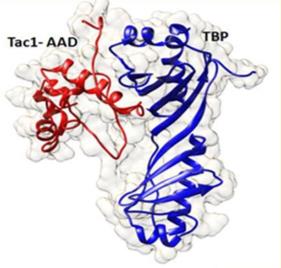 Fig. 1 Model depicting the interaction of TAC1-AAD and TBP.1
Key structural properties of tac1:
- Typical random flexible conformation with a neuropeptide precursor
- The C-terminal region contains a highly conserved receptor-binding motif
- Characteristic FFGLM five peptide sequence constitutes active center
- Phenylalanine (7) and methionine (8) play a decisive role in receptor activation
Functions of tac1
The main function of the Tac1 gene is to encode the neuropeptide P substance, which acts as an endogenous ligand of the neurokinin receptor and participates in various physiological processes. In addition, it also involves neurogenic inflammation, pain signal transduction and stress response regulation.
| Function | Description |
| Pain signal transduction | Substance P transmits and amplifies pain signals by activating the NK1 receptor, especially playing a key role in chronic pain. |
| Neurogenic inflammation | Mediates vasodilation and plasma extravasation, connecting the nervous system with inflammatory responses. |
| Regulation of emotional stress | Regulate anxiety-like behaviors and stress responses in brain regions such as the amygdala. |
| Regulation of gastrointestinal motility | As a neurotransmitter of the intestinal nervous system, it affects the peristalsis and secretory functions of the gastrointestinal tract. |
| Regulation of cell proliferation | Signal transduction pathways through influence the growth of a particular cell differentiation process. |
Unlike the multifunctional properties of most neuropeptides, other members of the tachykinin family, such as neurokinin A, are mainly involved in smooth muscle contraction. This functional specificity stems from the unique receptor selectivity and distribution pattern of substance P, making it an important molecular bridge connecting sensory nerves and the immune system.
Applications of tac1 and tac1 Antibody in Literature
1. He, Zi-Xuan, et al. "A nucleus accumbens Tac1 neural circuit regulates avoidance responses to aversive stimuli." International Journal of Molecular Sciences 24.5 (2023): 4346. https://doi.org/10.3390/ijms24054346
Research has found that Tac1 neurons in the medial shell of the nucleus accumbens can sense aversive stimuli and drive avoidance behaviors. It regulates this motivational behavior through the Tac1→ lateral hypothalamus pathway and by receiving excitatory inputs from the prefrontal cortex.
2. Lloyd, Elliot, et al. "Tac1 Deficiency Reduces the Severity of Enteric Bacterial Infection." bioRxiv (2025). https://doi.org/10.1101/2025.05.27.656414
Research has found that in Citrobacter rodents infection, the intestinal pathogen load in Tac1 gene-deficient mice is significantly reduced, the inflammatory response is weakened, and the recruitment of specific immune cells to the colon is decreased, indicating that sensory neuropeptides can regulate the host's immune response to intestinal infection.
3. Jain, Tushar, et al. "Molecular dissection studies of TAC1, a transcription activator of Candida drug resistance genes of the human pathogenic fungus Candida albicans." Frontiers in Microbiology 14 (2023): 994873. https://doi.org/10.3389/fmicb.2023.994873
Research has found that the transcription factor Tac1p of Candida albicans regulates the expression of the ABC transporter by binding to the CDR gene promoter at the N-terminal, recruiting TBP at the C-terminal, and potentially serving as an exogenous binding domain in the middle section, thereby driving multidrug resistance to azole drugs.
4. Xu, Hai, et al. "LOC134466 methylation promotes oncogenesis of endometrial carcinoma through LOC134466/hsa-miR-196a-5p/TAC1 axis." Aging (albany NY) 10.11 (2018): 3353. https://doi.org/10.18632/aging.101644
Studies have found that in endometrial cancer, highly methylated LOC134466 upregates TAC1 by adsorbing hsa-miR-196a-5p, thereby activating the neuroactive ligand-receptor interaction pathway and ultimately promoting tumor progression.
5. Chen, Yu‐Ling, et al. "Phox2a in Lateral Spinal Nucleus Tac1‐Positive Neurons Mediates Histamine‐Independent Acute Itch." CNS Neuroscience & Therapeutics 31.11 (2025): e70639.https://doi.org/10.1111/cns.70639
Research has found that neurons expressing Tac1 in the lateral nucleus of the spinal cord dominate histamine-independent pruritus. Phox2a within it inhibits pruritus by regulating presynaptic excitability, providing a new target for the treatment of this type of pruritus.
Creative Biolabs: tac1 Antibodies for Research
Creative Biolabs specializes in the production of high-quality tac1 antibodies for research and industrial applications. Our portfolio includes monoclonal antibodies tailored for ELISA, Flow Cytometry, Western blot, immunohistochemistry, and other diagnostic methodologies.
- Custom tac1 Antibody Development: Tailor-made solutions to meet specific research requirements.
- Bulk Production: Large-scale antibody manufacturing for industry partners.
- Technical Support: Expert consultation for protocol optimization and troubleshooting.
- Aliquoting Services: Conveniently sized aliquots for long-term storage and consistent experimental outcomes.
For more details on our tac1 antibodies, custom preparations, or technical support, contact us at email.
Reference
- Jain, Tushar, et al. "Molecular dissection studies of TAC1, a transcription activator of Candida drug resistance genes of the human pathogenic fungus Candida albicans." Frontiers in Microbiology 14 (2023): 994873. https://doi.org/10.3389/fmicb.2023.994873
Anti-tac1 antibodies
 Loading...


Hot products 
-
Mouse Anti-ENO1 Recombinant Antibody (CBYC-A950) (CBMAB-A4388-YC)

-
Mouse Anti-EIF4G1 Recombinant Antibody (2A9) (CBMAB-A2544-LY)

-
Mouse Anti-ALB Recombinant Antibody (V2-363290) (CBMAB-S0173-CQ)

-
Mouse Anti-CDKL5 Recombinant Antibody (CBFYC-1629) (CBMAB-C1689-FY)

-
Mouse Anti-B2M Recombinant Antibody (CBYY-0050) (CBMAB-0050-YY)

-
Mouse Anti-ABL2 Recombinant Antibody (V2-179121) (CBMAB-A0364-YC)

-
Mouse Anti-AAV9 Recombinant Antibody (V2-634029) (CBMAB-AP023LY)

-
Mouse Anti-FOXL1 Recombinant Antibody (CBXF-0845) (CBMAB-F0462-CQ)

-
Mouse Anti-ATP1B1 Recombinant Antibody (E4) (CBMAB-0463-LY)

-
Mouse Anti-CHRNA9 Recombinant Antibody (8E4) (CBMAB-C9161-LY)

-
Mouse Anti-BAX Recombinant Antibody (CBYY-0216) (CBMAB-0217-YY)

-
Mouse Anti-ABIN2 Recombinant Antibody (V2-179106) (CBMAB-A0349-YC)

-
Mouse Anti-BrdU Recombinant Antibody (IIB5) (CBMAB-1038CQ)

-
Mouse Anti-ADRB2 Recombinant Antibody (V2-180026) (CBMAB-A1420-YC)

-
Rabbit Anti-CBL Recombinant Antibody (D4E10) (CBMAB-CP0149-LY)

-
Mouse Anti-CTNND1 Recombinant Antibody (CBFYC-2414) (CBMAB-C2487-FY)

-
Mouse Anti-COL1A2 Recombinant Antibody (CF108) (V2LY-1206-LY626)

-
Rabbit Anti-ALK (Phosphorylated Y1278) Recombinant Antibody (D59G10) (PTM-CBMAB-0035YC)

-
Mouse Anti-AAV-5 Recombinant Antibody (V2-503417) (CBMAB-V208-1369-FY)

-
Mouse Anti-BRD3 Recombinant Antibody (CBYY-0801) (CBMAB-0804-YY)

- AActivation
- AGAgonist
- APApoptosis
- BBlocking
- BABioassay
- BIBioimaging
- CImmunohistochemistry-Frozen Sections
- CIChromatin Immunoprecipitation
- CTCytotoxicity
- CSCostimulation
- DDepletion
- DBDot Blot
- EELISA
- ECELISA(Cap)
- EDELISA(Det)
- ESELISpot
- EMElectron Microscopy
- FFlow Cytometry
- FNFunction Assay
- GSGel Supershift
- IInhibition
- IAEnzyme Immunoassay
- ICImmunocytochemistry
- IDImmunodiffusion
- IEImmunoelectrophoresis
- IFImmunofluorescence
- IGImmunochromatography
- IHImmunohistochemistry
- IMImmunomicroscopy
- IOImmunoassay
- IPImmunoprecipitation
- ISIntracellular Staining for Flow Cytometry
- LALuminex Assay
- LFLateral Flow Immunoassay
- MMicroarray
- MCMass Cytometry/CyTOF
- MDMeDIP
- MSElectrophoretic Mobility Shift Assay
- NNeutralization
- PImmunohistologyp-Paraffin Sections
- PAPeptide Array
- PEPeptide ELISA
- PLProximity Ligation Assay
- RRadioimmunoassay
- SStimulation
- SESandwich ELISA
- SHIn situ hybridization
- TCTissue Culture
- WBWestern Blot
