Cell Activation
Flow cytometry is normally combined with highly specific fluorophore-conjugated antibodies that will only bind to the activated forms of molecules. As the technology develops, advances in flow cytometry enable to perform quantitative multiplexed analysis of single cells within heterogeneous populations stained with specific antibodies for phenotyping in conjunction with antibodies to phosphorylated such as activated molecules within signaling pathways. By reactivating signaling pathways in vitro, it is easy to collect data on the responsive state of complex cell populations such as immune cells.
T Cell Activation
T cell activation requires at least two signals to become fully activated. The first occurs after engagement of the T cell antigen-specific receptor (TCR) by the antigen-major histocompatibility complex (MHC). The second occurs by subsequent engagement of co-stimulatory molecules such as CD28. These signals are transmitted to the nucleus and result in:
- Clonal expansion of T cells
- Upregulation of activation markers on the cell surface
- Differentiation into effector cells
- Induction of cytotoxicity or cytokine secretion
- Induction of apoptosis
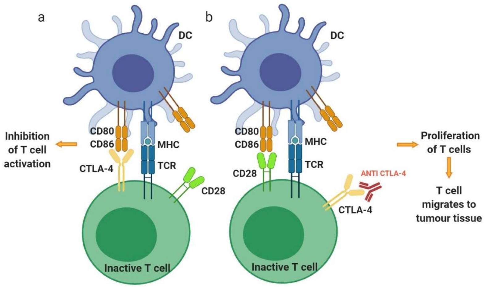 Fig.1 T cell activation1.
Before you begin your immune cell activation experiment, there are several points you need to take into consideration.
- Firstly, you should identify the cells of interest for your assays, whether that means isolating them from a mixed cell population or using cell surface markers to distinguish them from other cell types.
- Secondly, the method of stimulation you choose will impact how you measure the activation that occurs. For instance, a degranulation assay would be more appropriate if stimulating cells of interest with target cells, whereas antigenic and chemical stimulation would be preferential for proliferation studies.
- Lastly, be aware that activating immune cells can result in a change in phenotype, gene regulation, and function.
Here are three major ways to examine immune cell activation via flow cytometry.
- Surface Antibody Staining
- Intracellular Cytokine Staining
- Cell Proliferation
Before encountering a pathogen, immune cells exist in a resting state. Exposure to a pro-inflammatory milieu or stimulation with a foreign pathogen leads to an intracellular signaling cascade that culminates with genetic changes resulting in alterations to cell surface proteins. For example, when presented with the appropriate antigen, T cell will be stimulated to present maturation markers such as CD69 that aid in proliferation of this newly differentiated effector cell. Changes in the expression of surface proteins can be measured by flow cytometry.
Cytokine measurement allows researchers to investigate the response to specific antigen stimulation, and the use of flow cytometry provides the ability to determine what cytokines are being produced by what specific cell type
Following a response to a pathogen, immune cells will become activated and proliferate to increase their population numbers. This is an important point to study, particularly when trying to uncover what immune cell populations expand the most during a specific infection.
General activation protocols normally use pharmacological reagents and antibodies, making them ideal to determine immune competence, marker upregulation, cytokine release and proliferation by flow cytometry. Protocol of Cell Activation for Flow Cytometry is presented with the following parts:
- Unprimed T cell activation - Pharmacologic Method
- Unprimed T cell activation - Antibody Stimulation Method
- Primed T cell activation - Antigen Presenting Cell Co-culture
Please refer to the corresponding section according to your specific demands.
Reference
-
Alard, Emilie, et al. "Advances in anti-cancer immunotherapy: Car-T cell, checkpoint inhibitors, dendritic cell vaccines, and oncolytic viruses, and emerging cellular and molecular targets." Cancers 12.7 (2020): 1826.
Distributed under Open Access license CC BY 4.0, without modification.