RECK Antibodies
Background
The RECK protein (Reversion inducing Cysteine Protein, with Kazal motifs) is a membrane bound glycoprotein that is predominantly found in endothelial cells in the body. It plays a role in controlling the activity of matrix metalloproteinases (MMP) which're enzymes that break down proteins in the extracellular matrix to help maintain tissue structure and stability. Apart from its involvement, in cell adhesion and movement and tissue healing processes RECK also acts to suppress tumor growth by blocking cancer cells from invading surrounding tissues and spreading to parts of the body. Since its discovery, in 1997 the study of RECK has garnered attention in the fields of cancer biology and tissue balance regulation as it unveils understandings of disease processes and promising treatment avenues.
Structure of RECK
RECK is a transmembrane glycoprotein with a molecular weight of approximately 50 kDa. This weight may vary slightly among different species due to differences in glycosylation patterns or minor variations in amino acid sequences.
| Species | Human | Mouse | Rabbit | Chicken |
| Molecular Weight (kDa) | 50 | 50 | 50 | 50 |
| Primary Structural Differences | Contains multiple Kazal-like motifs and a transmembrane domain. The protein is highly conserved with critical cysteine residues involved in disulfide bonding. | Highly conserved with human RECK, sharing similar Kazal-like motifs and transmembrane domain. Minor differences may exist in non-critical regions. | Structure is highly conserved with human RECK, but may have slight differences in glycosylation patterns. | Contains conserved Kazal-like motifs, but may have some differences in the transmembrane domain and non-critical regions compared to mammalian RECK. |
The protein RECK contains about 400 amino acids which forms a transmembrane structure with multiple domains. The primary structure of RECK contains several Kazal-like motifs which are cysteine-rich regions that enable its inhibitory action against matrix metalloproteinases (MMPs). The transmembrane domain of RECK functions as a membrane anchor that keeps the protein attached to cell membranes. The protein structure consists of separate domains where the Kazal-like motifs located in the extracellular region bind to MMPs. The transmembrane domain maintains RECK's stable position in cell membranes which enables the protein to control MMP activity and preserve extracellular matrix structure. The Kazal-like motifs of RECK contain essential functional residues which enable its inhibitory function while the protein structure maintains its role as a vital tissue remodeling and cell behavior regulator.
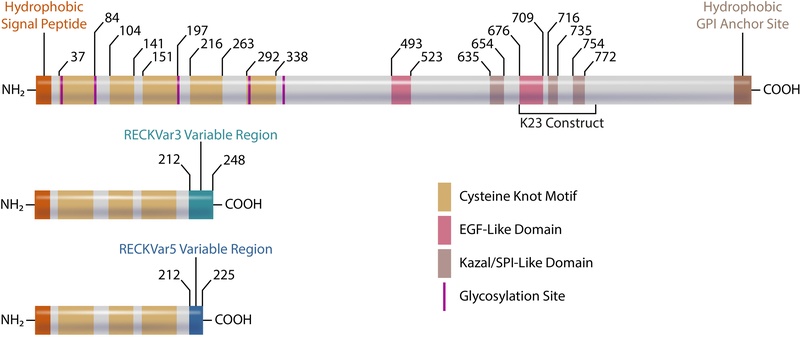 Fig. 1 Domains/motifs of canonical RECK (top) and RECK isoforms.1
Key structural properties of RECK:
- Transmembrane domain anchors it to the cell membrane
- Multiple Kazal-like motifs inhibit matrix metalloproteinases
- GPI anchor at the C-terminus ensures membrane attachment
- Cysteine knot motifs in the N-terminus involved in angiogenesis.
- EGF-like domains with potential roles in ligand shedding.
- Serine protease inhibitor sites regulate protease activity.
Functions of RECK
RECKs main role is to block matrix metalloproteinases (or MMP enzymes) which help preserve the structure of the cell environment known as the matrix. Additionally, RECK plays roles, in functions, like cell sticking adhesion) movement (migration) and keeping tumors in check.
| Function | Description |
| Tumor Suppression | Prevents tumor invasion and metastasis by regulating MMP activity. |
| Cell Adhesion | Promotes cell adhesion and maintains tissue integrity. |
| Angiogenesis | Enhances oxygen availability in tissues during low-oxygen conditions, supporting survival in hypoxic environments. |
| Blood-Brain Barrier Maintenance | Contributes to blood vessel formation and development. |
| Inhibition of MMPs | RECK regulates the activity of matrix metalloproteinases, preventing excessive degradation of the extracellular matrix. |
| Wnt Signaling Modulation | Involved in modulating Wnt signaling pathways, influencing cell fate and differentiation. |
The role of RECK, in preventing MMP activity is crucial for preserving tissue structure and function differently from its involvement in supporting cell adhesion; emphasizing its role, in managing matrix strength and cell communication.
Applications of RECK and RECK Antibody in Literature
1. Kimura, Tokuhiro, et al. "RECK is up-regulated and involved in chondrocyte cloning in human osteoarthritic cartilage." The American journal of pathology 176.6 (2010): 2858-2867. https://doi.org/10.2353/ajpath.2010.091003
The article identifies RECK as a fundamental element for chondrocyte cloning in human osteoarthritic cartilage. The study shows that RECK expression increases in this condition while demonstrating its essential role in osteoarthritis development. The research offers essential knowledge about cartilage degeneration mechanisms which could lead to new therapeutic approaches that target RECK for osteoarthritis treatment.
2. Mahl, Christian, et al. "RECK (reversion-inducing cysteine-rich protein with Kazal motifs) regulates migration, differentiation and Wnt/β-catenin signaling in human mesenchymal stem cells." Cellular and Molecular Life Sciences 73 (2016): 1489-1501. https://doi.org/10.1007/s00018-015-2054-4
The article delves into how RECK influences the behavior of mesenchymal stem cells (hMSCs). It has been observed that RECK plays a role in controlling cell movement and specialization as impacting Wnt/β catenin signaling within these cells.The results underscore the significance of RECK in regulating functions in hMSCs and hint at its possible use as a target, for treating conditions related to tissue healing and renewal.
3. Yoshida, Yoko, et al. "Suppression of tumor metastasis by a RECK-activating small molecule." Scientific Reports 12.1 (2022): 2319. https://doi.org/10.1038/s41598-022-06288-3
The article presents RECK as a crucial factor that limits tumor metastasis and describes a small molecule called DSK638 which enhances RECK expression. The RECK-activating compound shows strong potential to block metastasis in cancer models through its ability to increase RECK expression and subsequent downstream effects. The research indicates that DSK638 could become a valuable therapeutic tool for treating metastatic tumors by utilizing RECK's tumor-suppressive properties.
4. Matsuzaki, Tomoko, et al. "The RECK tumor-suppressor protein binds and stabilizes ADAMTS10." Biology open 7.10 (2018): bio033985. https://doi.org/10.1242/bio.033985
The article shows that RECK functions as a tumor suppressor and describes its relationship with ADAMTS10. The study shows that RECK binds to and stabilizes ADAMTS10 which functions as a tumor-suppressive metalloprotease. The binding between these proteins strengthens their anti-tumorigenic properties which could lead to new ways to stop tumor growth. The research indicates that blocking the RECK-ADAMTS10 pathway presents a promising therapeutic approach for cancer treatment.
5. Qi, Fangyuan, et al. "Identification of RECK as a protective prognostic indicator and a tumor suppressor through regulation of the ERK/MAPK signaling pathway in gastric cancer." Journal of Translational Medicine 21.1 (2023): 766. https://doi.org/10.1186/s12967-023-04644-z
The article establishes RECK as a vital factor in gastric cancer development and shows its dual function as a protective prognostic indicator and tumor suppressor. The study shows that RECK controls the ERK/MAPK signaling pathway which plays a vital role in cancer cell growth and survival. The tumor-suppressive effects of RECK occur through its regulation of this pathway which makes it a promising biomarker for prognosis and therapeutic target in gastric cancer.
Creative Biolabs: RECK Antibodies for Research
Creative Biolabs develops and manufactures innovative RECK-related products which serve both research and therapeutic purposes. Our product range consists of RECK antibodies with high binding affinity as well as recombinant RECK proteins and RECK-activating compounds which help researchers study cancer biology and metastasis suppression and tissue repair. Our products are designed to serve researchers and clinicians who want to improve disease understanding and treatment of conditions influenced by RECK.
- Custom RECK Antibody Development: We offer tailor-made RECK antibody solutions to meet specific research needs.
- Bulk Production: We provide large-scale manufacturing of RECK antibodies for industrial partners.
- Technical Support: Our team of experts offers consultation for protocol optimization and troubleshooting.
- Aliquoting Services: We provide conveniently sized aliquots to ensure long-term stability and consistent results in your experiments.
For further information on our RECK antibodies, custom orders, or technical assistance, please reach out to us via info@creative-biolabs.com.
Reference
- Russell, Jacob J., et al. "Reversion inducing cysteine rich protein with Kazal motifs and cardiovascular diseases: The RECKlessness of adverse remodeling." Cellular signalling 83 (2021): 109993. https://doi.org/10.1016/j.cellsig.2021.109993
Anti-RECK antibodies
 Loading...


Hot products 
-
Mouse Anti-AKT1 Recombinant Antibody (V2-180546) (CBMAB-A2070-YC)

-
Rat Anti-(1-5)-α-L-Arabinan Recombinant Antibody (V2-501861) (CBMAB-XB0003-YC)

-
Mouse Anti-CTNND1 Recombinant Antibody (CBFYC-2414) (CBMAB-C2487-FY)

-
Mouse Anti-F11R Recombinant Antibody (402) (CBMAB-0026-WJ)

-
Mouse Anti-ALDOA Recombinant Antibody (A2) (CBMAB-A2316-YC)

-
Mouse Anti-CD46 Recombinant Antibody (CBFYC-0076) (CBMAB-C0085-FY)

-
Mouse Anti-EPO Recombinant Antibody (CBFYR0196) (CBMAB-R0196-FY)

-
Mouse Anti-BBS2 Recombinant Antibody (CBYY-0253) (CBMAB-0254-YY)

-
Mouse Anti-NSUN6 Recombinant Antibody (D-5) (CBMAB-N3674-WJ)

-
Mouse Anti-BMI1 Recombinant Antibody (CBYC-P026) (CBMAB-P0108-YC)

-
Mouse Anti-APP Recombinant Antibody (5C2A1) (CBMAB-A3314-YC)

-
Mouse Anti-CCDC6 Recombinant Antibody (CBXC-0106) (CBMAB-C5397-CQ)

-
Rabbit Anti-ADRA1A Recombinant Antibody (V2-12532) (CBMAB-1022-CN)

-
Mouse Anti-CD24 Recombinant Antibody (ALB9) (CBMAB-0176CQ)

-
Mouse Anti-CORO1A Recombinant Antibody (4G10) (V2LY-1206-LY806)

-
Rabbit Anti-BAD (Phospho-Ser136) Recombinant Antibody (CAP219) (CBMAB-AP536LY)

-
Mouse Anti-CD2AP Recombinant Antibody (BR083) (CBMAB-BR083LY)

-
Mouse Anti-GIPC2 Recombinant Antibody (10) (CBMAB-G0476-LY)

-
Mouse Anti-CRTAM Recombinant Antibody (CBFYC-2235) (CBMAB-C2305-FY)

-
Mouse Anti-CAT Recombinant Antibody (724810) (CBMAB-C8431-LY)

- AActivation
- AGAgonist
- APApoptosis
- BBlocking
- BABioassay
- BIBioimaging
- CImmunohistochemistry-Frozen Sections
- CIChromatin Immunoprecipitation
- CTCytotoxicity
- CSCostimulation
- DDepletion
- DBDot Blot
- EELISA
- ECELISA(Cap)
- EDELISA(Det)
- ESELISpot
- EMElectron Microscopy
- FFlow Cytometry
- FNFunction Assay
- GSGel Supershift
- IInhibition
- IAEnzyme Immunoassay
- ICImmunocytochemistry
- IDImmunodiffusion
- IEImmunoelectrophoresis
- IFImmunofluorescence
- IGImmunochromatography
- IHImmunohistochemistry
- IMImmunomicroscopy
- IOImmunoassay
- IPImmunoprecipitation
- ISIntracellular Staining for Flow Cytometry
- LALuminex Assay
- LFLateral Flow Immunoassay
- MMicroarray
- MCMass Cytometry/CyTOF
- MDMeDIP
- MSElectrophoretic Mobility Shift Assay
- NNeutralization
- PImmunohistologyp-Paraffin Sections
- PAPeptide Array
- PEPeptide ELISA
- PLProximity Ligation Assay
- RRadioimmunoassay
- SStimulation
- SESandwich ELISA
- SHIn situ hybridization
- TCTissue Culture
- WBWestern Blot
