TRADD Antibodies
Background
TRADD (Tumor necrosis factor receptor type 1-associated death domain protein), as a key adaptor protein, is mainly present in the cytoplasm of mammals. This protein binds to the tumor necrosis factor receptor through its death domain and participates in mediating apoptosis, inflammatory responses, and the activation of the NF-κB signaling pathway, thereby maintaining immune homeostasis. Under conditions of viral infection or cellular stress, TRADD regulates the balance between cell survival and death by forming different signaling complexes. This gene was first identified by Hsu et al. in 1995. Its unique bridging function provides a key breakthrough for revealing the signal transduction mechanism of death receptors. The context-specific role of TRADD molecules in multiple signaling pathways remains a key model in the research of programmed cell death and immune regulation to this day, greatly promoting the development of therapeutic strategies for cancer and autoimmune diseases.
Structure of TRADD
TRADD is a adaptor protein with a molecular weight of approximately 34 kDa. Its precise molecular weight varies slightly among different species, mainly due to minor variations in amino acid sequences.
| Species | Human | Mouse | Rat | Bovine | Rhesus monkey |
| Molecular Weight (kDa) | 34.0 | 33.8 | 33.9 | 34.2 | 33.9 |
| Primary Structural Differences | Highly conservative death domains | The homology of death domains is high | Highly similar to human TRADD function | There is a slight variation at the carboxyl terminal | Highly consistent with the human protein sequence |
In the human body, it mainly expresses a highly conserved Death Domain (DD), which is responsible for interacting with members of the TNF receptor superfamily and is a key mediator in apoptosis and the NF-κB signaling pathway.
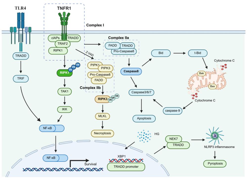 Fig. 1 TRADD-mediated signaling pathways.1
Key structural properties of TRADD:
- Contains a C-terminal death domain
- The N-terminal area is relatively loose
- Charge distribution on the surface of the dead domain
- Lack of inherent enzymatic activity
Functions of TRADD
The core function of the TRADD protein is to serve as a key adaptor protein for the signal transduction of tumor necrosis factor receptor 1 (TNFR1). However, it is also widely involved in regulating a variety of crucial cellular processes such as apoptosis, necroptosis, inflammatory responses and cell survival.
| Function | Description |
| Apoptotic signal guide | Through the recruitment of FADD and Caspase-8 by its Death Domain, the death inducing signaling complex (DISC) is formed to initiate the apoptosis program. |
| Activation of the NF-κB pathway | As a platform protein, it recruits molecules such as RIPK1 and TRAF2, activates the NF-κB signaling pathway, promotes the expression of pro-survival and inflammatory genes, and inhibits apoptosis. |
| Regulation of necrotic apoptosis | When the activity of Caspase-8 is inhibited, the complex involved in TRADD can turn to activate the RIPK3/MLKL pathway, inducing necrotic apoptosis. |
| Inflammatory response mediation | By activating the NF-κB pathway and inducing the production of inflammatory factors such as interleukins, it plays a core role in the body's immune defense. |
| Cell fate decision-making | Depending on the differences in cell types and microenvironmental signals, it dynamically participates in the formation of different signal complexes and is a key molecular switch that determines the survival or death of cells. |
The signal transduction of TRADD does not rely on enzyme activity. Its function is entirely determined by its structural characteristics as a molecular "platform" or "bridge", dynamically assembling different signal complexes through protein-protein interactions, thereby precisely regulating distinct cellular fates.
Applications of TRADD and TRADD Antibody in Literature
1. Sun, Kai, et al. "Inhibition of TRADD ameliorates chondrocyte necroptosis and osteoarthritis by blocking RIPK1-TAK1 pathway and restoring autophagy." Cell Death Discovery 9.1 (2023): 109. https://doi.org/10.1038/s41420-023-01406-0
This study reveals that TRADD mediates TNF-α -induced necrotic apoptosis of chondrocytes and osteoarthritis phenotype. Inhibiting TRADD can block the RIPK1-TAK1-NF-κB signaling and restore autophagy, thereby alleviating cartilage degeneration. The TRADD selective inhibitor ICCB-19 has demonstrated therapeutic potential both in vivo and in vitro, providing a potential therapeutic strategy for OA.
2. Wang, Xinyue, et al. "Rescue RM/CS-AKI by blocking strategy with one-dose anti-myoglobin RabMAb." Nature Communications 16.1 (2025): 1044. https://doi.org/10.1038/s41467-019-08584-5
This study reveals that TRADD plays a key role in the survival of mice in the context of RIPK1/RIPK3 deletion. The absence of TRADD can partially save the perinatal mortality of Ripk1−/−Ripk3−/− mice, but premature death still occurs in adulthood. TRADD plays a superior role in NFκB signaling compared to RIPK1 and promotes TNFα -induced thymocyte apoptosis.
3. Feoktistova, Maria, et al. "RIPK1 and TRADD regulate TNF-induced signaling and ripoptosome formation." International Journal of Molecular Sciences 22.22 (2021): 12459. https://doi.org/10.3390/ijms222212459
This study reveals the key role of TRADD in TNF signaling: it does not participate in necroptosis, but is essential for apoptosis and may negatively regulate NIK stability to inhibit ripoptosome formation. The research also found that RIPK1 can prevent the TNF-induced degradation of TRADD, and the two work together to regulate the balance of cell life and death.
4. Wang, Lili, et al. "TRADD mediates RIPK1-independent necroptosis induced by tumor necrosis factor." Frontiers in cell and developmental biology 7 (2020): 393. https://doi.org/10.3389/fcell.2019.00393
This study reveals that in RIPK1 knockdown cells, TRADD is a key factor in TNF-induced necrotic apoptosis. TRADD activates the RIPK3-MLKL signaling pathway and induces ROS accumulation by binding to RIPK3 to form a complex, promoting RIPK3 oligomerization and phosphorylation, and ultimately mediates necrotic apoptosis independently of RIPK1.
5. Koo, Gi-Bang, et al. "Nuclear TRADD prevents DNA damage-mediated death by facilitating non-homologous end-joining repair." Scientific reports 7.1 (2017): 3332. https://doi.org/10.1038/s41598-017-03211-z
This study reveals that TRADD translocations from the cytoplasm to the double-strand break point in the DNA damage response, promoting the repair of non-homologous terminal joins by recruiting 53BP1 and Ku70/80 complexes, and maintaining genomic stability. The absence of TRADD nuclear loci will lead to repair disorders, continuous activation of JNK and accumulation of ROS, ultimately causing cell death.
Creative Biolabs: TRADD Antibodies for Research
Creative Biolabs specializes in the production of high-quality TRADD antibodies for research and industrial applications. Our portfolio includes monoclonal antibodies tailored for ELISA, Flow Cytometry, Western blot, immunohistochemistry, and other diagnostic methodologies.
- Custom TRADD Antibody Development: Tailor-made solutions to meet specific research requirements.
- Bulk Production: Large-scale antibody manufacturing for industry partners.
- Technical Support: Expert consultation for protocol optimization and troubleshooting.
- Aliquoting Services: Conveniently sized aliquots for long-term storage and consistent experimental outcomes.
For more details on our TRADD antibodies, custom preparations, or technical support, contact us at email.
Reference
- Wang, Xueling, et al. "The Dual Role of TRADD in Liver Disease: From Cell Death Regulation to Inflammatory Microenvironment Remodeling." International Journal of Molecular Sciences 26.12 (2025): 5860. https://doi.org/10.3390/ijms26125860
Anti-TRADD antibodies
 Loading...


Hot products 
-
Mouse Anti-AAV9 Recombinant Antibody (V2-634029) (CBMAB-AP023LY)

-
Mouse Anti-AQP2 Recombinant Antibody (E-2) (CBMAB-A3358-YC)

-
Mouse Anti-BANF1 Recombinant Antibody (3F10-4G12) (CBMAB-A0707-LY)

-
Mouse Anti-ATM Recombinant Antibody (2C1) (CBMAB-A3970-YC)

-
Mouse Anti-CD59 Recombinant Antibody (CBXC-2097) (CBMAB-C4421-CQ)

-
Rat Anti-CCR2 Recombinant Antibody (475301) (CBMAB-C1338-LY)

-
Mouse Anti-ADGRE5 Recombinant Antibody (V2-360335) (CBMAB-C2088-CQ)

-
Mouse Anti-ADRB2 Recombinant Antibody (V2-180026) (CBMAB-A1420-YC)

-
Mouse Anti-APOH Recombinant Antibody (4D9A4) (CBMAB-A3249-YC)

-
Mouse Anti-CECR2 Recombinant Antibody (CBWJC-2465) (CBMAB-C3533WJ)

-
Mouse Anti-AQP2 Recombinant Antibody (G-3) (CBMAB-A3359-YC)

-
Mouse Anti-4-Hydroxynonenal Recombinant Antibody (V2-502280) (CBMAB-C1055-CN)

-
Mouse Anti-Acetyl-α-Tubulin (Lys40) Recombinant Antibody (V2-623485) (CBMAB-CP2897-LY)

-
Mouse Anti-CCNH Recombinant Antibody (CBFYC-1054) (CBMAB-C1111-FY)

-
Mouse Anti-BCL6 Recombinant Antibody (CBYY-0435) (CBMAB-0437-YY)

-
Mouse Anti-DLL4 Recombinant Antibody (D1090) (CBMAB-D1090-YC)

-
Mouse Anti-APOE Recombinant Antibody (A1) (CBMAB-0078CQ)

-
Rat Anti-EPO Recombinant Antibody (16) (CBMAB-E1578-FY)

-
Mouse Anti-ANXA7 Recombinant Antibody (A-1) (CBMAB-A2941-YC)

-
Mouse Anti-C1QC Recombinant Antibody (CBFYC-0600) (CBMAB-C0654-FY)

- AActivation
- AGAgonist
- APApoptosis
- BBlocking
- BABioassay
- BIBioimaging
- CImmunohistochemistry-Frozen Sections
- CIChromatin Immunoprecipitation
- CTCytotoxicity
- CSCostimulation
- DDepletion
- DBDot Blot
- EELISA
- ECELISA(Cap)
- EDELISA(Det)
- ESELISpot
- EMElectron Microscopy
- FFlow Cytometry
- FNFunction Assay
- GSGel Supershift
- IInhibition
- IAEnzyme Immunoassay
- ICImmunocytochemistry
- IDImmunodiffusion
- IEImmunoelectrophoresis
- IFImmunofluorescence
- IGImmunochromatography
- IHImmunohistochemistry
- IMImmunomicroscopy
- IOImmunoassay
- IPImmunoprecipitation
- ISIntracellular Staining for Flow Cytometry
- LALuminex Assay
- LFLateral Flow Immunoassay
- MMicroarray
- MCMass Cytometry/CyTOF
- MDMeDIP
- MSElectrophoretic Mobility Shift Assay
- NNeutralization
- PImmunohistologyp-Paraffin Sections
- PAPeptide Array
- PEPeptide ELISA
- PLProximity Ligation Assay
- RRadioimmunoassay
- SStimulation
- SESandwich ELISA
- SHIn situ hybridization
- TCTissue Culture
- WBWestern Blot
