
IFNAR1 Antibodies
Background
IFNAR1, as a key component of the type I interferon receptor, is mainly expressed on the surface of various cells in vertebrates. The transmembrane protein encoded by this gene can specifically bind to type I interferons and thereby activate the downstream JAK-STAT signaling pathway, thereby activating the cell's antiviral and immune regulatory functions. During the body's response to viral infection, IFNAR1 coordinates the innate immune response by maintaining the stability of the interferon signaling pathway. Its functional deficiency can lead to a decline in immune defense capabilities. This gene was fully identified in the early 1990s, and the analysis of its receptor complex structure has deepened our understanding of the cytokine signal transduction mechanism. Its rigorous multi-domain composition and dynamic regulatory mechanism have become an important model for the study of immune receptors, providing a key theoretical basis for the treatment of autoimmune diseases and the development of antiviral drugs.
Structure of IFNAR1
IFNAR1 is a transmembrane protein with a molecular weight of approximately 60 kDa. The exact value may vary slightly among different species due to glycosylation modifications and differences in amino acid sequences.
| Species | Human | Mouse | Rhesus monkey | Rat |
| Molecular Weight (kDa) | ~60 | ~61 | ~60 | ~61 |
| Primary Structural Differences | Extracellular region contains four fibronectin III structure domain, is a key area of ligand binding | With the human high homology, intracellular signal transduction structure domain highly conservative | The sequence is highly similar to that of humans and is commonly used in preclinical research. | Across the membrane area highly conservative, guarantee the receptor to anchor |
This protein is composed of approximately 557 amino acids. Its structural feature is that a single transmembrane region divides the molecule into an extracellular ligand-binding domain and an intracellular signal-transduction domain. The extracellular domain forms a rigid ligand-binding interface through multiple β-sheet structures, while the intracellular domain itself has no kinase activity, but contains conserved Box1/Box2 motifs, which are used to couple JAK kinases (mainly Tyk2), thereby initiating the phosphorylation and nuclear translocation signal cascade of downstream STAT proteins.
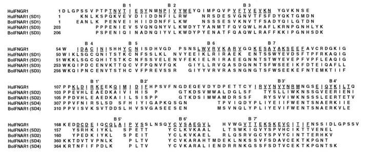 Fig. 1 Alignment of human IFNGR-1 with bovine and human IFNAR-1 sequences.1
Key structural properties of IFNAR1:
- Multidomain extracellular region
- Single transmembrane helix
- Conservative intracellular motif
- No intrinsic kinase activity
Functions of IFNAR1
The core function of the IFNAR1 gene is to mediate the signal transduction of type I interferons, thereby activating the innate immune response of cells. Additionally, it is involved in regulating various physiological and pathological processes such as immune balance, cell proliferation, and differentiation.
| Function | Description |
| Signal Transduction Initiation | The IFNAR1 and IFNAR2 together form a receptor complex. After binding to type I interferons (such as IFN-α/β), they activate the associated JAK kinases (Tyk2 and JAK1). |
| Immune response activation | The activated JAK kinases phosphorylate STAT1/STAT2 proteins, forming the transcriptional complex ISGF3, which then enters the cell nucleus and initiates the expression of interferon-stimulated genes (ISGs), establishing an antiviral state. |
| Immune Homeostasis Regulation | It regulates the intensity and duration of signals through negative feedback mechanisms, preventing excessive immune responses and participating in the pathological processes of autoimmune diseases. |
| Cell Proliferation and Apoptosis Effects | In certain cell types, continuous signal activation can induce cell cycle arrest or apoptosis, exerting anti-tumor effects. |
| Development and Differentiation Involvement | Studies in mouse models have shown that these signaling pathways play a regulatory role in the development, differentiation, and functional maturation of immune cells. |
Unlike many receptors with enzymatic activity, IFNAR1 itself does not possess catalytic function. Its signal transduction is entirely dependent on the cascade reaction with JAK kinases and STAT transcription factors, which reflects its characteristic as a crucial "hub" rather than an "effector" in the immune signaling network.
Applications of IFNAR1 and IFNAR1 Antibody in Literature
1. Cutrone, Elizabeth Cali, and Jerome A. Langer. "Identification of critical residues in bovine IFNAR-1 responsible for interferon binding." Journal of Biological Chemistry 276.20 (2001): 17140-17148. https://doi.org/10.1074/jbc.M009663200
The article indicates that type I interferon in humans functions through the IFNAR receptor, with the IFNAR-1 subunit playing a crucial role in binding specificity. The study utilized a more affinity-matched bovine IFNAR-1 model to identify five aromatic residues on its surface as forming the ligand binding site. This discovery is consistent with the functional studies of the human receptor and reveals a novel binding topological structure.
2. Wan, Qianya, et al. "PIM1 Attenuates Innate Immunity to Foster Coronavirus Replication through Ubiquitin Ligase β‐TrCP‐Mediated IFNAR1 Degradation." Advanced Science 12.37 (2025): e03487. https://doi.org/10.1002/advs.202503487
The research has found that the novel coronavirus HCoV-OC43 enhances the ubiquitination and degradation of the interferon receptor IFNAR1 by upregulating the host protein PIM1 kinase, thereby weakening the innate immunity. Inhibiting the activity of PIM1 can effectively block this process and suppress viral replication, providing a new target for the development of antiviral drugs.
3. Sun, Yimin, et al. "IFNAR1 gene mutation may contribute to developmental stuttering in the Chinese population." Hereditas 158.1 (2021): 46. https://doi.org/10.1186/s41065-021-00211-y
This study conducted whole-exome sequencing on families with speech disorders and for the first time discovered that variations in the IFNAR1 gene are associated with developmental stuttering. This gene mutation impairs the activity of the type I interferon signaling pathway, suggesting a potential pathogenic mechanism in the onset of stuttering.
4. Li, Huangcan, et al. "Secreted LRPAP1 binds and triggers IFNAR1 degradation to facilitate virus evasion from cellular innate immunity." Signal transduction and targeted therapy 8.1 (2023): 374. https://doi.org/10.1038/s41392-023-01630-1
The research has found that the proteases of the novel coronavirus SARS-CoV-2 and the enterovirus EV71 can upregulate the secretion of the LRPAP1 protein. This protein binds to and degrades the interferon receptor IFNAR1, thereby helping the virus evade the immune system. Inhibiting this pathway can stabilize IFNAR1 and effectively suppress various viruses.
5. Hussain, Timon, et al. "IFNAR1 deficiency impairs immunostimulatory properties of neutrophils in tumor-draining lymph nodes." Frontiers in immunology 13 (2022): 878959. https://doi.org/10.3389/fimmu.2022.878959
The research has found that in the lymph nodes draining head and neck cancer tumors, the absence of the function of type I interferon receptor IFNAR1 leads to the accumulation of neutrophils, but it will weaken their interaction with T cells and activation ability, thereby promoting tumor growth. Activating this pathway can enhance the immune stimulating function of neutrophils and inhibit tumors.
Creative Biolabs: IFNAR1 Antibodies for Research
Creative Biolabs specializes in the production of high-quality IFNAR1 antibodies for research and industrial applications. Our portfolio includes monoclonal antibodies tailored for ELISA, Flow Cytometry, Western blot, immunohistochemistry, and other diagnostic methodologies.
- Custom IFNAR1 Antibody Development: Tailor-made solutions to meet specific research requirements.
- Bulk Production: Large-scale antibody manufacturing for industry partners.
- Technical Support: Expert consultation for protocol optimization and troubleshooting.
- Aliquoting Services: Conveniently sized aliquots for long-term storage and consistent experimental outcomes.
For more details on our IFNAR1 antibodies, custom preparations, or technical support, contact us at email.
Reference
- Cutrone, Elizabeth Cali, and Jerome A. Langer. "Identification of critical residues in bovine IFNAR-1 responsible for interferon binding." Journal of Biological Chemistry 276.20 (2001): 17140-17148. https://doi.org/10.1074/jbc.M009663200
Anti-IFNAR1 antibodies
 Loading...
 Compare




Hot products 
-
Rabbit Anti-B2M Recombinant Antibody (CBYY-0059) (CBMAB-0059-YY)

-
Mouse Anti-GLP1R Recombinant Antibody (4F3) (CBMAB-G0521-LY)

-
Mouse Anti-APCS Recombinant Antibody (CBYC-A663) (CBMAB-A3054-YC)

-
Mouse Anti-GPRC5C Recombinant Antibody (CBFYH-0464) (CBMAB-H0521-FY)

-
Mouse Anti-CD46 Recombinant Antibody (CBFYC-0076) (CBMAB-C0085-FY)

-
Mouse Anti-CHRNA9 Recombinant Antibody (8E4) (CBMAB-C9161-LY)

-
Mouse Anti-AOC3 Recombinant Antibody (CBYY-0014) (CBMAB-0014-YY)

-
Mouse Anti-EGR1 Recombinant Antibody (CBWJZ-100) (CBMAB-Z0289-WJ)

-
Mouse Anti-ASTN1 Recombinant Antibody (H-9) (CBMAB-1154-CN)

-
Mouse Anti-ADGRE5 Recombinant Antibody (V2-360335) (CBMAB-C2088-CQ)

-
Mouse Anti-ASH1L Monoclonal Antibody (ASH5H03) (CBMAB-1372-YC)

-
Mouse Anti-GFAP Recombinant Antibody (24) (CBMAB-G2927-LY)

-
Mouse Anti-ACE2 Recombinant Antibody (V2-179293) (CBMAB-A0566-YC)

-
Mouse Anti-AKT1 Recombinant Antibody (V2-180546) (CBMAB-A2070-YC)

-
Rabbit Anti-CBL Recombinant Antibody (D4E10) (CBMAB-CP0149-LY)

-
Mouse Anti-ARG1 Recombinant Antibody (CBYCL-103) (CBMAB-L0004-YC)

-
Mouse Anti-ACVR1C Recombinant Antibody (V2-179685) (CBMAB-A1041-YC)

-
Mouse Anti-Gro Monoclonal Antibody (1) (CBMAB-0482-LY)

-
Mouse Anti-CDK7 Recombinant Antibody (CBYY-C1783) (CBMAB-C3221-YY)

-
Mouse Anti-CAPZB Recombinant Antibody (CBYY-C0944) (CBMAB-C2381-YY)

- AActivation
- AGAgonist
- APApoptosis
- BBlocking
- BABioassay
- BIBioimaging
- CImmunohistochemistry-Frozen Sections
- CIChromatin Immunoprecipitation
- CTCytotoxicity
- CSCostimulation
- DDepletion
- DBDot Blot
- EELISA
- ECELISA(Cap)
- EDELISA(Det)
- ESELISpot
- EMElectron Microscopy
- FFlow Cytometry
- FNFunction Assay
- GSGel Supershift
- IInhibition
- IAEnzyme Immunoassay
- ICImmunocytochemistry
- IDImmunodiffusion
- IEImmunoelectrophoresis
- IFImmunofluorescence
- IGImmunochromatography
- IHImmunohistochemistry
- IMImmunomicroscopy
- IOImmunoassay
- IPImmunoprecipitation
- ISIntracellular Staining for Flow Cytometry
- LALuminex Assay
- LFLateral Flow Immunoassay
- MMicroarray
- MCMass Cytometry/CyTOF
- MDMeDIP
- MSElectrophoretic Mobility Shift Assay
- NNeutralization
- PImmunohistologyp-Paraffin Sections
- PAPeptide Array
- PEPeptide ELISA
- PLProximity Ligation Assay
- RRadioimmunoassay
- SStimulation
- SESandwich ELISA
- SHIn situ hybridization
- TCTissue Culture
- WBWestern Blot
