
DOK2 Antibodies
Background
The DOK2 gene encodes a adaptor protein, which is mainly present in hematopoietic cells and serves as a key regulatory factor in signal transduction. This protein interacts with various signaling molecules through its phosphotyrosine binding domain, participates in regulating the development, activation and function of immune cells, and plays an important role in maintaining immune homeostasis. DOK2 was first discovered in the 1990s and attracted attention due to its binding to ABL oncoprotein in chronic myeloid leukemia. Subsequent studies have gradually revealed its negative regulatory functions in signaling pathways such as T-cell receptors and B-cell receptors. Its structure is relatively simple, but it has multiple regulatory interfaces, providing an important model for studying the fine regulation of immune signal networks and the molecular mechanisms of cell proliferation and differentiation. It is particularly significant in research related to autoimmune diseases and hematological malignancies.
Structure of DOK2
DOK2 is a adaptor protein with a molecular weight of approximately 48 kDa. Its molecular weight may vary under different isomers or post-translational modification states.
| Species | Human | Mouse | Rat |
| Molecular Weight (kDa) | 48 | 47.5 | 48.2 |
| Primary Structural Differences | Containing an N-terminal PH domain and a PTB domain | High homology with human, functional conservation | Highly similar core structure domain |
The DOK2 protein is composed of 476 amino acids and presents a typical structure of a signal adaptor protein as a whole. Its primary structure consists of the N-terminal pleckstrin homologous (PH) domain and the phosphotyrosine binding (PTB) domain, the latter of which is crucial for recognizing and binding phosphorylated tyrosine residues. The C-terminal region is rich in proline and multiple phosphorylation sites, which can serve as a docking platform for the SH2 domain. This structure enables DOK2 to connect with the activation signal linkers on the cell membrane through the PTB domain and recruit downstream effector molecules through the C-terminal, thereby playing a negative regulatory role in receptor signal transduction.
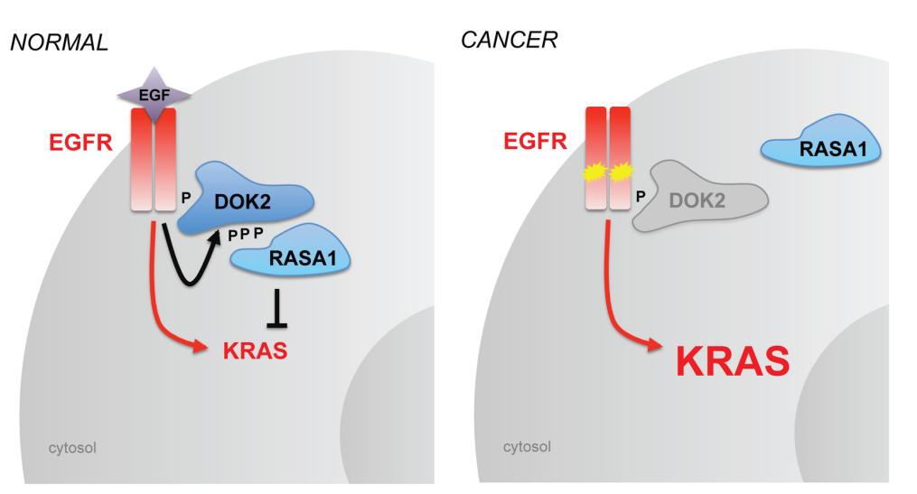 Fig. 1 Model of DOK2 function in normal lung and lung adenocarcinoma.1
Key structural properties of DOK2:
- Multi-domain assembly mode
- PTB domain structure formed tyrosine phosphate combined with pockets
- C-terminal region is rich in proline and phosphorylable sites
- Structural conformational changes regulate its inhibitory function as a "connector" for signal transduction
Functions of DOK2
The core function of the DOK2 protein is to serve as a negative regulatory hub in signal transduction. However, it is also involved in various physiological and pathological processes such as cell proliferation, differentiation, and the balance of immune responses.
| Function | Description |
| Negative regulation of signal transduction | Recruited downstream of immune cell receptors, they attenuate activation signals by recruiting inhibitory proteins or enzymes. |
| Maintenance of immune homeostasis | By negatively regulating the activation thresholds of T cells, B cells, etc., it prevents excessive immune responses and maintains autoimmune tolerance. |
| Cell proliferation inhibition | In certain environments, it can inhibit the proliferation signal driven by growth factors, and its functional inactivation is related to the occurrence and development of some hematological malignancies. |
| Participation in the apoptotic process | By participating in the regulation of specific signaling pathways, it influences the survival and apoptosis decisions of cells. |
| Differentiation support | In hematopoietic cell lineage differentiation such as regulating role in the process, affect the generation of functional cells. |
The mode of action of DOK2 is similar to that of a "molecular damper", and its inhibitory effect is gradual and continuous, which is quite different from some signal molecules that act instantaneously, demonstrating its unique role in precisely maintaining the steady state of signal pathways.
Applications of DOK2 and DOK2 Antibody in Literature
1. Xu, Jiaxuan, et al. "DOK2 Has Prognostic and Immunologic Significance in Adults With Acute Myeloid Leukemia: A Novel Immune-Related Therapeutic Target." Frontiers in Medicine 9 (2022): 842383. https://doi.org/10.3389/fmed.2022.842383
This study reveals that the DOK2 gene is significantly upregulated in acute myeloid leukemia (AML), and high expression suggests shorter overall survival and disease-free survival for patients. Functional analysis revealed that DOK2 is involved in immune regulation and can serve as a potential biomarker for the poor prognosis of AML.
2. Berger, Alice H., et al. "DOK2 inhibits EGFR-mutated lung adenocarcinoma." PLoS One 8.11 (2013): e79526. https://doi.org/10.1371/journal.pone.0079526
This study confirmed that DOK2 is a tumor suppressor gene for EGFR-mutated lung adenocarcinoma. Its genomic deletion coexists with EGFR mutations, and it has been demonstrated through mouse models that Dok2 deletion accelerates the occurrence of EGFR-driven lung cancer. Mechanically, DOK2 is recruited by mutant EGFR and inhibits RAS activation, forming a negative feedback loop.
3. Laletin, Vladimir, et al. "DOK1 and DOK2 regulate CD8 T cell signaling and memory formation without affecting tumor cell killing." Scientific Reports 14.1 (2024): 15053. https://doi.org/10.1038/s41598-024-66075-0
This study focuses on T-cell signal suppressor proteins DOK1 and DOK2. It was found that knockout of DOK1/DOK2 could enhance the TCR signaling pathway activity of pre-stimulated CD8+ T cells and promote the effector memory phenotype, but its in vitro anti-melanoma ability did not significantly improve, suggesting that the DOK protein has an inhibitory effect on T cells in long-term activation.
4. Lyons, Anthony, et al. "Dok2 mediates the CD200Fc attenuation of Aβ-induced changes in glia." Journal of neuroinflammation 9.1 (2012): 107. https://doi.org/10.1186/1742-2094-9-107
This study reveals that the activation of CD200R can inhibit the activation of microglia and the release of inflammatory factors induced by β -amyloid protein (Aβ), and improve the damage of synaptic plasticity in the hippocampus.
5. Tengda, Li, Qian Cheng, and Sun Yi. "Identification of melanoma subsets based on DNA methylation sites and construction of a prognosis evaluation model." Journal of Oncology 2022.1 (2022): 6608650. https://doi.org/10.1155/2022/6608650
This study classified melanoma into seven subtypes based on DNA methylation characteristics and constructed a prognostic risk model. Among them, the expression of key genes such as DOK2 is significantly correlated with the prognosis of patients and the stage of tumors. Their low expression is associated with a poor prognosis, providing a new target for the diagnosis and treatment of melanoma.
Creative Biolabs: DOK2 Antibodies for Research
Creative Biolabs specializes in the production of high-quality DOK2 antibodies for research and industrial applications. Our portfolio includes monoclonal antibodies tailored for ELISA, Flow Cytometry, Western blot, immunohistochemistry, and other diagnostic methodologies.
- Custom DOK2 Antibody Development: Tailor-made solutions to meet specific research requirements.
- Bulk Production: Large-scale antibody manufacturing for industry partners.
- Technical Support: Expert consultation for protocol optimization and troubleshooting.
- Aliquoting Services: Conveniently sized aliquots for long-term storage and consistent experimental outcomes.
For more details on our DOK2 antibodies, custom preparations, or technical support, contact us at email.
Reference
- Berger, Alice H., et al. "DOK2 inhibits EGFR-mutated lung adenocarcinoma." PLoS One 8.11 (2013): e79526. https://doi.org/10.1371/journal.pone.0079526
Anti-DOK2 antibodies
 Loading...
 Compare




Hot products 
- AActivation
- AGAgonist
- APApoptosis
- BBlocking
- BABioassay
- BIBioimaging
- CImmunohistochemistry-Frozen Sections
- CIChromatin Immunoprecipitation
- CTCytotoxicity
- CSCostimulation
- DDepletion
- DBDot Blot
- EELISA
- ECELISA(Cap)
- EDELISA(Det)
- ESELISpot
- EMElectron Microscopy
- FFlow Cytometry
- FNFunction Assay
- GSGel Supershift
- IInhibition
- IAEnzyme Immunoassay
- ICImmunocytochemistry
- IDImmunodiffusion
- IEImmunoelectrophoresis
- IFImmunofluorescence
- IGImmunochromatography
- IHImmunohistochemistry
- IMImmunomicroscopy
- IOImmunoassay
- IPImmunoprecipitation
- ISIntracellular Staining for Flow Cytometry
- LALuminex Assay
- LFLateral Flow Immunoassay
- MMicroarray
- MCMass Cytometry/CyTOF
- MDMeDIP
- MSElectrophoretic Mobility Shift Assay
- NNeutralization
- PImmunohistologyp-Paraffin Sections
- PAPeptide Array
- PEPeptide ELISA
- PLProximity Ligation Assay
- RRadioimmunoassay
- SStimulation
- SESandwich ELISA
- SHIn situ hybridization
- TCTissue Culture
- WBWestern Blot
