HNF1A Antibodies
Background
The HNF1A gene encodes a liver-enriched transcription factor belonging to the homologous domain protein family. It is mainly expressed in the pancreas, liver, kidneys, and intestines. This protein regulates the expression of multiple target genes by binding to specific DNA sequences and plays a crucial role in glucose metabolism, lipid homeostasis, and pancreatic β-cell function. Mutations in the HNF1A gene are the most common cause of adult-onset type 3 diabetes (MODY3) in adolescents, leading to impaired insulin secretion and early-onset diabetes. The gene was isolated and identified in 1991 and was subsequently confirmed as the pathogenic gene for MODY. This discovery not only deepened our understanding of the pathogenesis of monogenic diabetes but also provided a model for the application of precision medicine in diabetes classification and individualized treatment. As a core node in the transcriptional regulatory network, the study of the structure and function of HNF1A continues to provide important insights into pancreatic development, metabolic regulation, and related disease mechanisms.
Structure of HNF1A
The protein encoded by the HNF1A gene has a molecular weight of approximately 67 kDa. There are slight variations among different species due to sequence differences. The specific data are as follows:
| Species | Human | Mouse | Rat | Pig | Cow |
| Molecular Weight (kDa) | 67.2 | 66.8 | 67.0 | 67.1 | 66.9 |
| Primary Structural Differences | Contains homologous domain | Highly homologous to humans | C-terminal variation | Moderately conserved | Functional domain conserved |
This protein is composed of 631 amino acids and contains three main domains: the N-terminal DNA-binding homologous domain, the middle POU domain, and the C-terminal trans-activating domain. Its spatial structure presents a typical transcription factor conformation. The POU domain is responsible for specific recognition of DNA sequences, while the homologous domain stabilizes the protein-DNA interaction. The protein binds to the promoter of the target gene by forming homodimers or heterodimers, regulating the expression of downstream genes. Phosphorylation modification can affect its transcriptional activity, and the nuclear localization signal ensures its entry into the cell nucleus to exert its function.
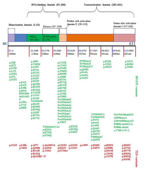 Fig. 1 The structure of HNF1α protein and the SNPs associated with MODY3 or T2D.1
Key structural features of the HNF1A gene:
- Encodes a transcription factor with a POU homology domain
- Dimerization domain mediates formation of homodimers or heterodimers
- DNA binding domain specifically recognizes the promoter sequence of target genes
- Transcriptional activation domain regulates the transcriptional activity of downstream genes
- Nuclear localization signal ensures the protein enters the nucleus to exert its function
- Phosphorylation sites participate in post-translational modification regulation
Functions of HNF1A
The core function of the HNF1A gene is to regulate the transcription of target genes in the pancreas, liver, and kidneys. However, it is also involved in various physiological processes, including glucose metabolism, lipid homeostasis, and cell differentiation.
| Function | Description |
| Blood sugar regulation | Activates insulin gene expression, maintains normal function of pancreatic β cells, and promotes glucose sensing. |
| Lipid metabolism regulation | Regulates the expression of apolipoproteins and lipid metabolism-related enzymes in the liver, affecting lipid homeostasis. |
| Kidney function | Regulates the expression of genes related to glucose reabsorption in the proximal tubules of the kidney. |
| Liver development | Participates in the differentiation of liver cells and the regulation of liver-specific gene expression. |
| Pancreas development | Maintains the characteristics of mature β cells, promotes normal development of the islets and maintenance of their functions. |
HNF1A forms homodimers or heterodimers by specifically binding to DNA. Its target genes include albumin, insulin, glucose transporters, and various liver enzymes. This regulatory network is crucial for maintaining metabolic homeostasis.
Applications of HNF1A and HNF1A Antibody in Literature
1. Li, Li-Mei, Bei-Ge Jiang, and Liang-Liang Sun. "HNF1A: from monogenic diabetes to type 2 diabetes and gestational diabetes mellitus." Frontiers in Endocrinology 13 (2022): 829565. https://doi.org/10.3389/fendo.2022.829565
The article indicates that mutations in the HNF1A gene can lead to three types of diabetes: MODY3, type 2 diabetes, and gestational diabetes. This gene is highly polymorphic, and the disease phenotypes and treatment methods triggered by different mutation sites vary. Analyzing its function is helpful for achieving precise classification and individualized treatment of diabetes.
2. Miyachi, Yasutaka, Takashi Miyazawa, and Yoshihiro Ogawa. "HNF1A mutations and beta cell dysfunction in diabetes." International journal of molecular sciences 23.6 (2022): 3222. https://doi.org/10.3390/ijms23063222
The article indicates that mutations in the HNF1A gene can cause monogenic diabetes MODY, and its polymorphism is also related to type 2 diabetes. The research, through genetic-modified mice and human stem cell models, revealed that HNF1A mainly causes diabetes by influencing the functions of pancreatic β cells and liver metabolism.
3. Mirshahi, Uyenlinh L., et al. "Reduced penetrance of MODY-associated HNF1A/HNF4A variants but not GCK variants in clinically unselected cohorts." The American Journal of Human Genetics 109.11 (2022): 2018-2028. https://doi.org/10.1016/j.ajhg.2022.09.014
The study found that pathogenic variations in the HNF1A, HNF4A and GCK genes are not uncommon in the population (approximately 1 in 1500). The diabetes penetrance of HNF1A varies depending on the diagnostic setting (32% - 98%), while GCK is nearly fully penetrant in all populations, suggesting that the genetic interpretation of different genes needs to take into account the clinical context.
4. Ng, Natasha Hui Jin, et al. "HNF4A and HNF1A exhibit tissue specific target gene regulation in pancreatic beta cells and hepatocytes." Nature communications 15.1 (2024): 4288. https://doi.org/10.1038/s41467-024-48647-w
This study used ChIP-Seq to map the genome-wide binding profiles of HNF4A and HNF1A in pancreatic and hepatic cells. It revealed the tissue-specific regulation of HNF4A and confirmed that HNF1A can bind and regulate the expression of GPR39 in pancreatic β cells, providing new resources for elucidating the functions of diabetes-related genes.
5. Xekouki, P., et al. "HNF1A gene mutations and premature ovarian failure (POF): evidence from a clinical paradigm combining MODY 3 and POF." Hormones 23.2 (2024): 345-350. https://doi.org/10.1007/s42000-024-00529-y
The research found that the HNF1A gene mutation not only causes MODY but also that its protein is expressed in both human fetal and adult ovaries. Based on this, the researchers hypothesized that HNF1A might be involved in ovarian development, and its mutation might be related to the onset of premature ovarian insufficiency (POF).
Creative Biolabs: HNF1A Antibodies for Research
Creative Biolabs specializes in the production of high-quality HNF1A antibodies for research and industrial applications. Our portfolio includes monoclonal and polyclonal antibodies tailored for ELISA, Flow Cytometry, Western blot, immunohistochemistry, and other diagnostic methodologies.
- Custom HNF1A Antibody Development: Tailor-made solutions to meet specific research requirements.
- Bulk Production: Large-scale antibody manufacturing for industry partners.
- Technical Support: Expert consultation for protocol optimization and troubleshooting.
- Aliquoting Services: Conveniently sized aliquots for long-term storage and consistent experimental outcomes.
For more details on our HNF1A antibodies, custom preparations, or technical support, contact us at email.
Reference
- Li, Li-Mei, Bei-Ge Jiang, and Liang-Liang Sun. "HNF1A: from monogenic diabetes to type 2 diabetes and gestational diabetes mellitus." Frontiers in Endocrinology 13 (2022): 829565. Distributed under Open Access license CC BY 4.0, without modification. https://doi.org/10.3389/fendo.2022.829565
Anti-HNF1A antibodies
 Loading...


Hot products 
-
Mouse Anti-DES Monoclonal Antibody (440) (CBMAB-AP1857LY)

-
Mouse Anti-FOXA3 Recombinant Antibody (2A9) (CBMAB-0377-YC)

-
Mouse Anti-CHRNA9 Recombinant Antibody (8E4) (CBMAB-C9161-LY)

-
Mouse Anti-ENO2 Recombinant Antibody (H14) (CBMAB-E1341-FY)

-
Mouse Anti-BACE1 Recombinant Antibody (61-3E7) (CBMAB-1183-CN)

-
Rabbit Anti-DLK1 Recombinant Antibody (9D8) (CBMAB-D1061-YC)

-
Mouse Anti-AMACR Recombinant Antibody (CB34A) (CBMAB-CA034LY)

-
Mouse Anti-CFL1 (Phospho-Ser3) Recombinant Antibody (CBFYC-1770) (CBMAB-C1832-FY)

-
Mouse Anti-2C TCR Recombinant Antibody (V2-1556) (CBMAB-0951-LY)

-
Rabbit Anti-BAD (Phospho-Ser136) Recombinant Antibody (CAP219) (CBMAB-AP536LY)

-
Mouse Anti-GLP1R Recombinant Antibody (4F3) (CBMAB-G0521-LY)

-
Rabbit Anti-ADRA1A Recombinant Antibody (V2-12532) (CBMAB-1022-CN)

-
Mouse Anti-ADGRE5 Recombinant Antibody (V2-360335) (CBMAB-C2088-CQ)

-
Rabbit Anti-AP2M1 (Phosphorylated T156) Recombinant Antibody (D4F3) (PTM-CBMAB-0610LY)

-
Mouse Anti-ALPL Antibody (B4-78) (CBMAB-1009CQ)

-
Mouse Anti-AAV-5 Recombinant Antibody (V2-503416) (CBMAB-V208-1402-FY)

-
Rat Anti-C5AR1 Recombinant Antibody (8D6) (CBMAB-C9139-LY)

-
Mouse Anti-CD33 Recombinant Antibody (P67.6) (CBMAB-C10189-LY)

-
Mouse Anti-BAD (Phospho-Ser136) Recombinant Antibody (CBYY-0138) (CBMAB-0139-YY)

-
Rabbit Anti-AKT3 Recombinant Antibody (V2-12567) (CBMAB-1057-CN)

- AActivation
- AGAgonist
- APApoptosis
- BBlocking
- BABioassay
- BIBioimaging
- CImmunohistochemistry-Frozen Sections
- CIChromatin Immunoprecipitation
- CTCytotoxicity
- CSCostimulation
- DDepletion
- DBDot Blot
- EELISA
- ECELISA(Cap)
- EDELISA(Det)
- ESELISpot
- EMElectron Microscopy
- FFlow Cytometry
- FNFunction Assay
- GSGel Supershift
- IInhibition
- IAEnzyme Immunoassay
- ICImmunocytochemistry
- IDImmunodiffusion
- IEImmunoelectrophoresis
- IFImmunofluorescence
- IGImmunochromatography
- IHImmunohistochemistry
- IMImmunomicroscopy
- IOImmunoassay
- IPImmunoprecipitation
- ISIntracellular Staining for Flow Cytometry
- LALuminex Assay
- LFLateral Flow Immunoassay
- MMicroarray
- MCMass Cytometry/CyTOF
- MDMeDIP
- MSElectrophoretic Mobility Shift Assay
- NNeutralization
- PImmunohistologyp-Paraffin Sections
- PAPeptide Array
- PEPeptide ELISA
- PLProximity Ligation Assay
- RRadioimmunoassay
- SStimulation
- SESandwich ELISA
- SHIn situ hybridization
- TCTissue Culture
- WBWestern Blot
