
Napsin A Antibodies
Background
Napsin A is an acidic aspartic protease mainly expressed in type II alveolar epithelial cells and renal tubular epithelial cells. This protein plays a crucial role in maintaining normal lung function and pulmonary surface tension stability by participating in the cleavage processing of surfactant protein B precursor. In the diagnosis of lung cancer, Napsin A is often used as a specific marker for lung adenocarcinoma, and its combined use with TTF-1 can significantly improve the accuracy of pathological diagnosis. This gene was discovered by a research team in lung tissue in 1998. The encoded protease is also involved in the regulatory pathway of the renin-angiotensin system. Its tissue-specific expression pattern and stable biological function have made it an important molecular target for respiratory system disease research, providing a key direction for the analysis of lung development, tumor differentiation, and related pathological mechanisms.
Structure of Napsin A
Napsin A is an acidic aspartic protease with a molecular weight of approximately 38 kDa. The molecular weight varies among different species, mainly due to subtle changes in the gene encoding sequence.
| Species | Human | Mouse | Rat |
| Molecular Weight (kDa) | ~38 | ~39 | ~37.5 |
| Primary Structural Differences | Lung adenocarcinoma markers, involved in the processing of surfactant protein B precursor | Expressed during lung development, and functionally highly homologous to that of humans | Protease activity is significantly expressed in the kidneys and secreted in the lungs |
This protein is composed of 420 amino acids and its primary structure folds to form a typical proteinase dimer domain. Its active center contains two highly conserved aspartic acid residues (Asp74 and Asp231), which are crucial for catalyzing the hydrolysis function. The protein's tertiary structure forms a hydrophilic active pocket that specifically recognizes and cleaves the specific amino acid sequence in the substrate protein. This precise substrate specificity enables it to play a key role in the biosynthesis of pulmonary surfactant and tumor-related pathways.
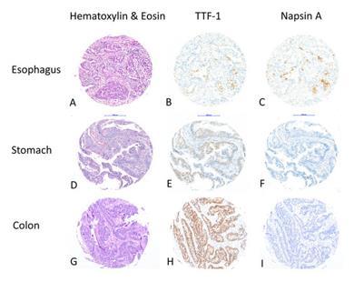 Fig. 1 Napsin A Expression in Gastrointestinal Adenocarcinomas.1
Key structural properties of Napsin A:
- Typical structure of a bifunctional aspartic protease
- The active center is composed of two conserved aspartic acid residues (Asp74, Asp231) to form a catalytic dimer
- Contains unique "flap" rings participate in identifying and combined with the substrate
- Precursor protein needs by removing n-terminal signal peptide and original structure domain to have an activity
Functions of Napsin A
The Napsin A gene encodes an aspartic acid protease. Its main function is to participate in the cleavage and maturation of the precursor of pulmonary surfactant protein B, which is crucial for maintaining the normal function of alveoli. Moreover, it also plays a role in various pathological processes.
| Function | Description |
| Pulmonary surfactant processing | It is specifically expressed in type II alveolar epithelial cells and is responsible for cleaving pro-SP-B into mature and active SP-B, which is essential for reducing alveolar surface tension and preventing lung collapse. |
| Tumor Diagnostic Markers | They are highly expressed in over 80% of lung adenocarcinomas and are important pathological diagnostic markers. They are often used in combination with TTF-1 to enhance diagnostic specificity and assist in differentiating primary lung adenocarcinoma from metastatic adenocarcinoma. |
| Tissue-specific expression | In addition to the lungs, it is also expressed in the distal tubules of the kidneys and the thyroid gland. However, the specific physiological function of this protein in the kidneys remains unclear. |
| Potential Pathophysiological Effects | The research suggests that abnormal expression of this protein may be associated with acute lung injury, pulmonary fibrosis, and certain kidney diseases. Its proteolytic activity may be involved in the regulation of the local tissue microenvironment. |
Unlike broad-spectrum aspartic proteases (such as cathepsin D), Napsin A has strict substrate specificity and tissue expression profiles, which determines its crucial role in specific physiological and pathological processes, rather than being a general protein degrading enzyme.
Applications of Napsin A and Napsin A Antibody in Literature
1. Noack, Petar, et al. "Immunohistochemical TTF-1 and Napsin a Expression in Gastrointestinal Adenocarcinomas—Low Frequency but an Important Pitfall." Diagnostics 15.12 (2025): 1490. https://doi.org/10.3390/diagnostics15121490
This study examined the expression of TTF-1 and Napsin A in 854 cases of gastrointestinal adenocarcinoma by immunohistochemistry. The results indicated that although this combination is commonly used for the diagnosis of lung adenocarcinoma, it is necessary to be aware of its abnormal expression in some gastrointestinal tumors to avoid misdiagnosis.
2. Li, Li, et al. "The high diagnostic accuracy of combined test of thyroid transcription factor 1 and Napsin A to distinguish between lung adenocarcinoma and squamous cell carcinoma: a meta-analysis." PLoS One 9.7 (2014): e100837. https://doi.org/10.1371/journal.pone.0100837
The article indicates that a meta-analysis, which combined data from 10 studies involving a total of 1,446 cases, found that the combined detection of TTF-1 and Napsin A can effectively distinguish lung adenocarcinoma from squamous cell carcinoma. The combined specificity reached 1.00, the sensitivity was 0.76, the area under the curve was 0.92, and it has significant diagnostic value.
3. Samukawa, Takuya, et al. "The elevation of serum napsin A in idiopathic pulmonary fibrosis, compared with KL-6, surfactant protein-A and surfactant protein-D." BMC pulmonary medicine 12.1 (2012): 55. https://doi.org/10.1186/1471-2466-12-55
This study suggests that serum Napsin A can serve as a potential biomarker for idiopathic pulmonary fibrosis. Compared with healthy controls, the serum Napsin A level in IPF patients is significantly elevated and is correlated with the severity of the disease. Its diagnostic efficacy may be superior to that of KL-6, SP-A, and SP-D.
4. Zhu, Bing, Stephen M. Rohan, and Xiaoqi Lin. "Immunoexpression of napsin A in renal neoplasms." Diagnostic pathology 10.1 (2015): 4. https://doi.org/10.1186/s13000-015-0242-z
This study examined the expression of Napsin A in 159 cases of renal tumors using tissue microarrays. The results showed that it had a relatively high positive rate in various subtypes of renal tumors (such as papillary renal cell carcinoma), indicating that this marker is not specific to lung adenocarcinoma. Therefore, when Napsin A is positive in metastatic cancer, differential diagnosis should also consider both lung and kidney origins.
5. Skirnisdottir, Ingiridur, et al. "Napsin A as a marker of clear cell ovarian carcinoma." BMC cancer 13.1 (2013): 524. https://doi.org/10.1186/1471-2407-13-524
The analysis of 131 cases of ovarian cancer in this study revealed that the positive rate of Napsin A in ovarian clear cell carcinoma was as high as 80%, significantly higher than that in other types of tumors. Combined with the expression status of p21 and p53, Napsin A can be used as an important auxiliary indicator for differentiating ovarian clear cell carcinoma.
Creative Biolabs: Napsin A Antibodies for Research
Creative Biolabs specializes in the production of high-quality Napsin A antibodies for research and industrial applications. Our portfolio includes monoclonal antibodies tailored for ELISA, Flow Cytometry, Western blot, immunohistochemistry, and other diagnostic methodologies.
- Custom Napsin A Antibody Development: Tailor-made solutions to meet specific research requirements.
- Bulk Production: Large-scale antibody manufacturing for industry partners.
- Technical Support: Expert consultation for protocol optimization and troubleshooting.
- Aliquoting Services: Conveniently sized aliquots for long-term storage and consistent experimental outcomes.
For more details on our Napsin A antibodies, custom preparations, or technical support, contact us at email.
Reference
- Noack, Petar, et al. "Immunohistochemical TTF-1 and Napsin a Expression in Gastrointestinal Adenocarcinomas—Low Frequency but an Important Pitfall." Diagnostics 15.12 (2025): 1490. Distributed under Open Access license CC BY 4.0, without modification.https://doi.org/10.3390/diagnostics15121490
Anti-Napsin A antibodies
 Loading...
 Compare




Hot products 
- AActivation
- AGAgonist
- APApoptosis
- BBlocking
- BABioassay
- BIBioimaging
- CImmunohistochemistry-Frozen Sections
- CIChromatin Immunoprecipitation
- CTCytotoxicity
- CSCostimulation
- DDepletion
- DBDot Blot
- EELISA
- ECELISA(Cap)
- EDELISA(Det)
- ESELISpot
- EMElectron Microscopy
- FFlow Cytometry
- FNFunction Assay
- GSGel Supershift
- IInhibition
- IAEnzyme Immunoassay
- ICImmunocytochemistry
- IDImmunodiffusion
- IEImmunoelectrophoresis
- IFImmunofluorescence
- IGImmunochromatography
- IHImmunohistochemistry
- IMImmunomicroscopy
- IOImmunoassay
- IPImmunoprecipitation
- ISIntracellular Staining for Flow Cytometry
- LALuminex Assay
- LFLateral Flow Immunoassay
- MMicroarray
- MCMass Cytometry/CyTOF
- MDMeDIP
- MSElectrophoretic Mobility Shift Assay
- NNeutralization
- PImmunohistologyp-Paraffin Sections
- PAPeptide Array
- PEPeptide ELISA
- PLProximity Ligation Assay
- RRadioimmunoassay
- SStimulation
- SESandwich ELISA
- SHIn situ hybridization
- TCTissue Culture
- WBWestern Blot
