
PLK1 Antibodies
Background
PLK1 is a highly conserved serine/threonine protein kinase, mainly present in eukaryotic cells, and plays a core regulatory role in the initiation, progression and completion of cell mitosis. This protein regulates key processes such as centrosome maturation, spindle assembly, chromosome separation and cytoplasmic division by phosphorylating multiple substrates, thereby maintaining genomic stability. Due to its significant role in the cell cycle, PLK1 has become a hot target in cancer research, and its overexpression is closely related to the proliferation and poor prognosis of various tumors. Since its discovery in the early 1990s, the structure and function of PLK1 have been resolved through X-ray crystallography and cryo-electron microscopy techniques, promoting the development of targeted inhibitors. Related research has significantly deepened people's understanding of the cell cycle regulation mechanism and targeted anti-cancer treatment strategies.
Structure of PLK1
PLK1 is a serine/threonine protein kinase with a molecular weight of approximately 68 kDa. The molecular weight of this protein varies slightly among different species, mainly due to the sequence diversity of its C-terminal domain.
| Species | Human | Mouse | African clawed toad | Fruit fly |
| Molecular Weight (kDa) | 68 | 67 | 66 | 65 |
| Primary Structural Differences | Contains the typical Polo box structure domain | Very high homology with humans | There are partial variations in the catalytic domain | The structure is relatively simplified |
The PLK1 protein is composed of 603 amino acids and forms a characteristic spatial conformation through its N-terminal kinase domain and C-terminal Polo box domain. The active center of this protein is located in the kinase domain, and the phosphorylation at the Thr210 site on its activation loop is the key to regulating enzyme activity. The Polo box domain, as a regulatory module, precisely guides the localization and functional performance of PLK1 within cells through its own conformational changes and interactions with other proteins, ensuring its spatiotemporal regulation of the mitotic process.
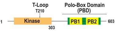 Fig. 1 Domain organization of PLK1.1
Key structural properties of PLK1:
- Modular structure of kinase domain and Polo box structure field synergy
- Activated ring segments in the kinase domain control catalytic activity
- Polo boxes constitute specific substrate recognition and subcellular localization modules
Functions of PLK1
The core function of PLK1 is to regulate the process of cell mitosis. In addition, this kinase is also involved in various cellular life activities such as DNA damage response, activation of cell cycle checkpoints, and regulation of cytoplasmic division.
| Function | Description |
| Mitosis initiation | Cells are pushed into the M phase by phosphorylated CDK1 activators, initiating the division program. |
| Centrosome maturation | Regulate the separation of centrosomes and the organization of microtubules to ensure the normal assembly of the spindle. |
| Chromosome separation | Participate in centromere function adjustment, ensure the chromatids separation accurately. |
| Cytoplasmic division | Guide the formation and positioning of the contraction ring to complete the final cell division. |
| DNA damage response | Pause the cycle process under the pressure of DNA replication to assist in the activation of the repair mechanism. |
The activity of PLK1 shows a strict cell cycle dependence, with its concentration reaching its peak at the G2/M phase. This dynamic change pattern is in perfect alignment with its functional positioning as the "main controller" of cell division, ensuring the precise coordination of division events in time and space.
Applications of PLK1 and PLK1 Antibody in Literature
1. Kalous, Jaroslav, and Daria Aleshkina. "Multiple roles of PLK1 in mitosis and meiosis." Cells 12.1 (2023): 187. https://doi.org/10.3390/cells12010187
The article indicates that PLK1, as a key cell cycle regulatory kinase, regulates the processes of mitosis and meiosis through phosphorylated substrates, affects spindle formation, chromosome segregation and other processes, and participates in the regulation of mRNA translation. At present, the specific mechanism of its action on meiosis remains to be further studied.
2. Du, Yang, et al. "Plk1 promotes renal tubulointerstitial fibrosis by targeting autophagy/lysosome axis." Cell Death & Disease 14.8 (2023): 571.https://doi.org/10.1038/s41419-023-06093-4
Research has found that PLK1 drives renal fibrosis in chronic kidney disease by regulating the autophagy/lysosomal axis. Inhibiting PLK1 can reduce autophagic flux, thereby suppressing fibroblast activation and kidney damage, indicating that it can serve as a new target for the treatment of chronic kidney disease.
3. Chiappa, Michela, et al. "Present and future perspective on PLK1 inhibition in cancer treatment." Frontiers in oncology 12 (2022): 903016. https://doi.org/10.3389/fonc.2022.903016
Research has found that PLK1 is a key kinase that regulates cell mitosis, DNA damage response and cell death. It is overexpressed in various tumors and promotes cancer through multiple pathways such as DNA repair and immunity, thus becoming a highly potential therapeutic target for anti-cancer treatment.
4. Ye, Peng, et al. "PLK1 inhibitors for the treatment of colorectal cancer." Annals of Medicine and Surgery 87.7 (2025): 4165-4172. https://doi.org/10.1097/MS9.0000000000003373
Research has found that PLK1 is an important target for anti-cancer treatment. Although the clinical efficacy of most PLK1 inhibitors is limited, the latest research indicates that they show potential for KRAS-mutated metastatic colorectal cancer, providing a new direction for research in this field.
5. Kim, Taekyung. "Recent Progress on the Localization of PLK1 to the Kinetochore and Its Role in Mitosis." International Journal of Molecular Sciences 23.9 (2022): 5252. https://doi.org/10.3390/ijms23095252
Research has found that PLK1, as a core kinase for cell division, regulates spindle assembly and chromosome separation through its precise localization on kinetosomes. This article mainly reviews the kinetogenic localization mechanism of PLK1 and its regulatory role in the key processes of mitosis.
Creative Biolabs: PLK1 Antibodies for Research
Creative Biolabs specializes in the production of high-quality PLK1 antibodies for research and industrial applications. Our portfolio includes monoclonal antibodies tailored for ELISA, Flow Cytometry, Western blot, immunohistochemistry, and other diagnostic methodologies.
- Custom PLK1 Antibody Development: Tailor-made solutions to meet specific research requirements.
- Bulk Production: Large-scale antibody manufacturing for industry partners.
- Technical Support: Expert consultation for protocol optimization and troubleshooting.
- Aliquoting Services: Conveniently sized aliquots for long-term storage and consistent experimental outcomes.
For more details on our PLK1 antibodies, custom preparations, or technical support, contact us at email.
Reference
- Kim, Taekyung. "Recent Progress on the Localization of PLK1 to the Kinetochore and Its Role in Mitosis." International Journal of Molecular Sciences 23.9 (2022): 5252. https://doi.org/10.3390/ijms23095252
Anti-PLK1 antibodies
 Loading...
 Compare




Hot products 
- AActivation
- AGAgonist
- APApoptosis
- BBlocking
- BABioassay
- BIBioimaging
- CImmunohistochemistry-Frozen Sections
- CIChromatin Immunoprecipitation
- CTCytotoxicity
- CSCostimulation
- DDepletion
- DBDot Blot
- EELISA
- ECELISA(Cap)
- EDELISA(Det)
- ESELISpot
- EMElectron Microscopy
- FFlow Cytometry
- FNFunction Assay
- GSGel Supershift
- IInhibition
- IAEnzyme Immunoassay
- ICImmunocytochemistry
- IDImmunodiffusion
- IEImmunoelectrophoresis
- IFImmunofluorescence
- IGImmunochromatography
- IHImmunohistochemistry
- IMImmunomicroscopy
- IOImmunoassay
- IPImmunoprecipitation
- ISIntracellular Staining for Flow Cytometry
- LALuminex Assay
- LFLateral Flow Immunoassay
- MMicroarray
- MCMass Cytometry/CyTOF
- MDMeDIP
- MSElectrophoretic Mobility Shift Assay
- NNeutralization
- PImmunohistologyp-Paraffin Sections
- PAPeptide Array
- PEPeptide ELISA
- PLProximity Ligation Assay
- RRadioimmunoassay
- SStimulation
- SESandwich ELISA
- SHIn situ hybridization
- TCTissue Culture
- WBWestern Blot
