
PSCA Antibodies
Background
PSCA is a cell surface glycoprotein belonging to the Thy-1/Ly-6 family and was initially identified in prostate basal cells. The protein encoded by this gene is anchored to the cell membrane through glycosylphosphatidylinositol (GPI), participating in cell signal transduction, adhesion and proliferation regulation, and is particularly expressed in the epithelial tissues of the urinary system and digestive tract. Research has found that PSCA is abnormally highly expressed in various cancers, such as prostate cancer, bladder cancer, and pancreatic cancer, and its expression level is closely related to tumor progression. Therefore, it has become an important target for immunotherapy and antibody drug development. Since its first report in 1998, the structural and functional studies of PSCA have provided a key molecular basis for tumor-targeted therapy strategies and promoted the clinical development of related monoclonal antibody drugs (such as AGS-1C4D4).
Structure of PSCA
PSCA is a cell surface glycoprotein with a molecular weight of approximately 12-15 kDa, and its precise molecular weight varies slightly depending on the degree of glycosylation. This protein is composed of 123 amino acids and has a typical GPI-anchored domain. Its core structure is a conserved three-finger-shaped fold of the Thy-1/Ly-6 family, composed of multiple β -folded sheets, and stabilized by disulfide bonds to maintain its receptor binding function. The following table lists the comparison of molecular characteristics of PSCA among different species:
| Species | Human | Mouse | Rat |
| Molecular Weight (kDa) | 12-15 | ~13 | ~13 |
| Primary Structural Differences | GPI anchored signal peptide was present | Highly homologous extracellular domain sequence | High homology with human |
The extracellular domain of PSCA contains a Ly-6/uPAR domain, which forms a specific spatial conformation through conserved disulfide bonds and is a key region for its binding to ligands or antibodies. The GPI-anchored signaling peptide at its C-terminal is removed during post-translational processing and the mature protein is fixed to the cell membrane.
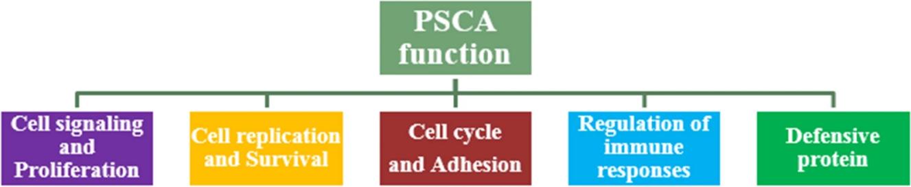 Fig. 1 Schematic view of PSCA function.1
Key structural properties of PSCA:
- Typical Ly-6/uPAR three-finger-shaped domain folding
- Maintain spatial conformational stability by relying on conserved disulfide bonds
- GPI anchoring modification mediates cell membrane localization
Functions of PSCA
The main function of the protein encoded by the PSCA gene is to participate in cell signal transduction and adhesion. However, its expression dysregulation is also closely related to many important pathological processes, especially playing a key role in tumorigenesis and development.
| Function | Description |
| Regulation of cell proliferation | By participating in specific signaling pathways (such as PI3K/Akt), it regulates the growth and survival of epithelial cells and maintains tissue homeostasis. |
| Intercellular adhesion | As a cell surface molecule, it affects the interactions between cells and between cells and the matrix. Its dysfunction may lead to enhanced cell migration. |
| Tumor promotion | In prostate cancer, bladder cancer and other cancers in the exceptionally high expression, such as by inhibiting apoptosis, promoting proliferation mechanism drive tumor progression. |
| Immunotherapy targets | Due to its specific membrane localization and high expression in tumor tissues, it has become an important target for immunotherapies such as antibody drugs and CAR-T. |
| Participation in developmental differentiation | In embryonic development and specific tissues, such as prostate, gastrointestinal tract play an important role in the process of cell differentiation. |
Unlike the relatively single high-affinity role of myosin in oxygen dynamics, the function of PSCA shows a high degree of background dependence, that is, it participates in maintaining homeostasis in normal tissues, but transforms into promoting malignant phenotypes in pathological states (especially in cancer), highlighting its potential and complexity as a therapeutic target.
Applications of PSCA and PSCA Antibody in Literature
1. Xu, Li-pu, et al. "Downregulation of PSCA promotes gastric cancer proliferation and is related to poor prognosis." Journal of Cancer 11.9 (2020): 2708. https://doi.org/10.7150/jca.33575
This study experimentally confirmed that the expression of prostate stem cell antigen (PSCA) was significantly down-regulated in gastric cancer tissues. Low expression of PSCA is associated with later TNM stage and poor prognosis, and it is an independent prognostic factor in multivariate analysis. Both in vitro and in vivo experiments have shown that PSCA can inhibit the proliferation of gastric cancer cells, suggesting its role as a tumor suppressor gene.
2. Usui, Yoshiaki, et al. "Impact of PSCA polymorphisms on the risk of duodenal ulcer." Journal of epidemiology 31.1 (2021): 12-20. https://doi.org/10.2188/jea.JE20190184
This study confirmed that in the Japanese population, the C allele at the rs2294008 locus of the PSCA gene significantly increases the risk of duodenal ulcer (DU), and the association is independent of environmental factors such as Helicobacter pylori infection. This site is not associated with the risk of gastric ulcer (GU).
3. Wu, Di, et al. "PSCA is a target of chimeric antigen receptor T cells in gastric cancer." Biomarker Research 8.1 (2020): 3. https://doi.org/10.1186/s40364-020-0183-x
In this study, third-generation CAR-T cells targeting PSCA were constructed, confirming that they could effectively activate and kill gastric cancer cells in vitro. Animal experiments have shown that local injection can inhibit tumor growth, but intravenous infusion has a poor therapeutic effect, suggesting that anti-PSCA CAR-T therapy has the potential for clinical transformation.
4. Dai, Zhenyu, et al. "Off-the-shelf invariant NKT cells expressing anti-PSCA CAR and IL-15 promote pancreatic cancer regression in mice." The Journal of Clinical Investigation 135.8 (2025). https://doi.org/10.1172/JCI179014
In this study, frozen off-the-shelf CAR-iNKT cells targeting PSCA were successfully developed. Experiments have confirmed that it can effectively inhibit pancreatic cancer, with a therapeutic effect comparable to that of CAR-T cells, and has not caused systemic toxicity such as graft-versus-host disease, showing a good clinical application prospect.
5. Nayerpour Dizaj, Tina, et al. "Significance of PSCA as a novel prognostic marker and therapeutic target for cancer." Cancer Cell International 24.1 (2024): 135. https://doi.org/10.1186/s12935-024-03320-6
The article indicates that prostate stem cell antigen (PSCA) is a functionally diverse cell surface protein in various cancers. Its expression level and genetic polymorphism have significant influences on cancer susceptibility and clinical characteristics. This review aims to systematically evaluate the alterations of PSCA in different cancer types and explore its clinical and therapeutic value as a diagnostic, prognostic and therapeutic target.
Creative Biolabs: PSCA Antibodies for Research
Creative Biolabs specializes in the production of high-quality PSCA antibodies for research and industrial applications. Our portfolio includes monoclonal antibodies tailored for ELISA, Flow Cytometry, Western blot, immunohistochemistry, and other diagnostic methodologies.
- Custom PSCA Antibody Development: Tailor-made solutions to meet specific research requirements.
- Bulk Production: Large-scale antibody manufacturing for industry partners.
- Technical Support: Expert consultation for protocol optimization and troubleshooting.
- Aliquoting Services: Conveniently sized aliquots for long-term storage and consistent experimental outcomes.
For more details on our PSCA antibodies, custom preparations, or technical support, contact us at email.
Reference
- Nayerpour Dizaj, Tina, et al. "Significance of PSCA as a novel prognostic marker and therapeutic target for cancer." Cancer Cell International 24.1 (2024): 135. https://doi.org/10.1186/s12935-024-03320-6
Anti-PSCA antibodies
 Loading...
 Compare




Hot products 
- AActivation
- AGAgonist
- APApoptosis
- BBlocking
- BABioassay
- BIBioimaging
- CImmunohistochemistry-Frozen Sections
- CIChromatin Immunoprecipitation
- CTCytotoxicity
- CSCostimulation
- DDepletion
- DBDot Blot
- EELISA
- ECELISA(Cap)
- EDELISA(Det)
- ESELISpot
- EMElectron Microscopy
- FFlow Cytometry
- FNFunction Assay
- GSGel Supershift
- IInhibition
- IAEnzyme Immunoassay
- ICImmunocytochemistry
- IDImmunodiffusion
- IEImmunoelectrophoresis
- IFImmunofluorescence
- IGImmunochromatography
- IHImmunohistochemistry
- IMImmunomicroscopy
- IOImmunoassay
- IPImmunoprecipitation
- ISIntracellular Staining for Flow Cytometry
- LALuminex Assay
- LFLateral Flow Immunoassay
- MMicroarray
- MCMass Cytometry/CyTOF
- MDMeDIP
- MSElectrophoretic Mobility Shift Assay
- NNeutralization
- PImmunohistologyp-Paraffin Sections
- PAPeptide Array
- PEPeptide ELISA
- PLProximity Ligation Assay
- RRadioimmunoassay
- SStimulation
- SESandwich ELISA
- SHIn situ hybridization
- TCTissue Culture
- WBWestern Blot
