
RELN Antibodies
Background
The RELN gene encodes a secretory glycoprotein called Reelin, which is mainly distributed in specific neurons in regions such as the cerebral cortex and hippocampus of mammals. Reelin plays a core role in the hierarchical structure of the embryonic cerebral cortex by regulating neuronal migration and precise localization. Mutations in this gene can lead to abnormal arrangement of neurons, which is closely related to various neurological disorders, such as autism spectrum disorder and schizophrenia. Since its discovery in 1995, the Reelin signaling pathway has become a key model in neurodevelopmental research, greatly advancing the understanding of cerebral cortex formation, synaptic plasticity, and related disease mechanisms.
Structure of RELN
The Reelin protein encoded by the RELN gene is a secreted macromolecular glycoprotein with a molecular weight of approximately 400 kDa. This protein is highly conserved in vertebrates.
| Species | Human | Mouse | Rat |
| Full-length amino acid quantity | 3460 | 3461 | 3460 |
| Core functional domain | Homologous structure and rodents, conservative function | Highly similar structures | Retain 8 EGF repeat sequences and 3 Reelin repeat units (central region) |
| Main functional differences | Gene mutations are associated with the risk of neuropsychiatric disorders such as schizophrenia and autism | Classic model animal, marked cortical inversion after knockout | Commonly used in electrical physiological and behavioral science research |
The primary structure of the Reelin protein consists of a signal peptide, an F-spondin domain, eight epidermal growth factor (EGF) repeat sequences, and three Reelin repeat units (RLD) rich in cationic amino acids at the C-terminal. Its tertiary structure is linearly arranged in series with multiple domains. The central RLD region is a key site for binding to its receptors (such as ApoER2 and VLDLR) and activating downstream signaling pathways. This protein is crucial for the formation of a normal layered structure in the cerebral cortex by regulating neuronal migration and synaptic plasticity.
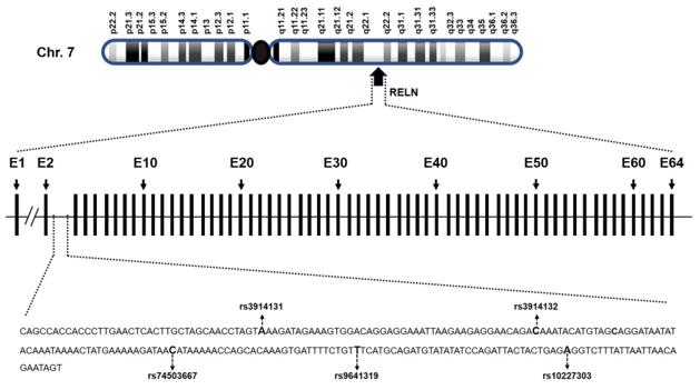 Fig. 1 Genomic location and organization of RELN gene.1
Key structural properties of RELN:
- Macromolecular secretory glycoproteins
- Repetitive functional domain
- Cationic binding region
Functions of RELN
The core function of the RELN gene is to regulate the migration and stratification of neurons in the cerebral cortex during the embryonic period, and it also participates in the regulation of synaptic plasticity in the adult nervous system.
| Function | Description |
| Guide neuron migration | The Reelin protein is secreted by Cajal-Retzius cells in the cerebral cortex, forming a concentration gradient that guides newly formed neurons to migrate correctly along the radial glial fibers to the target cortex. |
| Maintain the hierarchical structure of the cortex | Activating the downstream Dab1 signaling pathway and regulating the precise localization and stay of neurons at the migration endpoint is the key to the formation of a clear "inside-out" six-layer structure in the cerebral cortex. |
| Regulate synaptic plasticity | In the adult hippocampus and other brain regions continue to express, affect dendritic spines shape, NMDA receptor function and long range enhancement (LTP), involved in learning and memory process. |
| Signal pathway regulation | Combining ApoER2 and activate the cell membrane receptor and VLDLR, inducing intracellular cohesion Dab1 protein tyrosine phosphorylation, which affect the cytoskeleton rearrangement and gene expression. |
| Associated with diseases | Gene mutations or down-regulation of expression are associated with a variety of neuropsychiatric disorders, such as autism (abnormal cortical microstructure), schizophrenia (synaptic dysfunction), and Alzheimer's disease (Tau pathologically related). |
Unlike most signaling molecules that respond linearly or threshold, the function of Reelin depends on its local concentration gradient and spatiotemporal specific expression. This pattern enables it to precisely guide cell localization during development and play a continuous modulation role in mature neural circuits.
Applications of RELN and RELN Antibody in Literature
- Priyadarshi, Saurabh, et al. "The risks of RELN polymorphisms and its expression in the development of otosclerosis." Plos one 17.6 (2022): e0269558. https://doi.org/10.1371/journal.pone.0269558
Studies have confirmed that the RELN gene rs3914132 and rs74503667 variations significantly increase the risk of otosclerosis in different populations (including the Indian cohort) by regulating the binding of transcription factors, reducing gene and protein expression, and altering the morphology of the stapes.
- Li, Zhenpeng, et al. "Identification and validation of RELN mutation as a response indicator for immune checkpoint inhibitor therapy in melanoma and non-small cell lung cancer." Cells 11.23 (2022): 3841. https://doi.org/10.3390/cells11233841
Studies have shown that RELN gene mutations are associated with a better immune microenvironment, which can significantly increase the response rate and survival benefits of patients with melanoma and non-small cell lung cancer to immune checkpoint inhibitor treatment, and have predictive potential.
- Bracher-Smith, Matthew, et al. "Whole genome analysis in APOE4 homozygotes identifies the DAB1-RELN pathway in Alzheimer's disease pathogenesis." Neurobiology of Aging 119 (2022): 67-76. https://doi.org/10.1016/j.neurobiolaging.2022.07.009
Research has found that in the APOE-ε4 homozygous elderly population, the DAB1 locus (rs112437613) is significantly associated with the risk of Alzheimer's disease, and there is an upper-level genetic interaction between the DAB1-RELN signaling pathway and APOE.
- Ovadia, Galit, and Sagiv Shifman. "The genetic variation of RELN expression in schizophrenia and bipolar disorder." PloS one 6.5 (2011): e19955. https://doi.org/10.1371/journal.pone.0019955
Research has found that there are abnormalities in the RELN gene in schizophrenia and bipolar disorder: the proportion of short subtypes decreases, allele expression is imbalanced, and specific SNPS (rs7341475) are associated with the proportion of exon skipping in females.
- Arioka, Yuko, et al. "Single-cell trajectory analysis of human homogenous neurons carrying a rare RELN variant." Translational psychiatry 8.1 (2018): 129. https://doi.org/10.1038/s41398-018-0177-8
Research using iPSC technology has found that rare RELN gene mutations (RELN-DEL) can disrupt the Reelin signaling pathway, leading to impaired directional migration ability of human dopamine neurons and providing single-cell evidence for understanding the mechanism by which it causes mental illness.
Creative Biolabs: RELN Antibodies for Research
Creative Biolabs specializes in the production of high-quality RELN antibodies for research and industrial applications. Our portfolio includes monoclonal antibodies tailored for ELISA, Flow Cytometry, Western blot, immunohistochemistry, and other diagnostic methodologies.
- Custom RELN Antibody Development: Tailor-made solutions to meet specific research requirements.
- Bulk Production: Large-scale antibody manufacturing for industry partners.
- Technical Support: Expert consultation for protocol optimization and troubleshooting.
- Aliquoting Services: Conveniently sized aliquots for long-term storage and consistent experimental outcomes.
For more details on our RELN antibodies, custom preparations, or technical support, contact us at email.
Reference
- Priyadarshi, Saurabh, et al. "The risks of RELN polymorphisms and its expression in the development of otosclerosis." Plos one 17.6 (2022): e0269558. https://doi.org/10.1371/journal.pone.0269558
Anti-RELN antibodies
 Loading...
 Compare




Hot products 
- AActivation
- AGAgonist
- APApoptosis
- BBlocking
- BABioassay
- BIBioimaging
- CImmunohistochemistry-Frozen Sections
- CIChromatin Immunoprecipitation
- CTCytotoxicity
- CSCostimulation
- DDepletion
- DBDot Blot
- EELISA
- ECELISA(Cap)
- EDELISA(Det)
- ESELISpot
- EMElectron Microscopy
- FFlow Cytometry
- FNFunction Assay
- GSGel Supershift
- IInhibition
- IAEnzyme Immunoassay
- ICImmunocytochemistry
- IDImmunodiffusion
- IEImmunoelectrophoresis
- IFImmunofluorescence
- IGImmunochromatography
- IHImmunohistochemistry
- IMImmunomicroscopy
- IOImmunoassay
- IPImmunoprecipitation
- ISIntracellular Staining for Flow Cytometry
- LALuminex Assay
- LFLateral Flow Immunoassay
- MMicroarray
- MCMass Cytometry/CyTOF
- MDMeDIP
- MSElectrophoretic Mobility Shift Assay
- NNeutralization
- PImmunohistologyp-Paraffin Sections
- PAPeptide Array
- PEPeptide ELISA
- PLProximity Ligation Assay
- RRadioimmunoassay
- SStimulation
- SESandwich ELISA
- SHIn situ hybridization
- TCTissue Culture
- WBWestern Blot
