
TCRB Antibodies
Background
The TCRB gene is located on the long arm of human chromosome 7 (7q34), encoding the β chain of T-cell receptors, and is a core component of adaptive immune responses. This gene generates extremely rich TCR diversity through the V-D-J rearrangement mechanism, enabling T cells to recognize multiple pathogens. The β chain and α chain encoded by it jointly form the TCR complex, which specifically binds to the MHC-peptide complex during the antigen presentation process. This gene was first cloned by the team led by Tak Mak in 1984. It was the first T-cell receptor gene to be fully analyzed. This breakthrough directly promoted the development of tumor immunotherapy (such as CAR-T technology) and the research on the mechanism of autoimmune diseases. The continuous analysis of the TCRB gene structure not only reveals the molecular basis of lymphocyte development, but also provides a key theoretical basis for the development of diagnostic markers and the design of targeted therapies for immune-related diseases.
Structure of TCRB
The T-cell receptor β chain encoded by the TCRB gene is a transmembrane protein with a molecular weight of approximately 32-34 kDa, and the specific value varies depending on the degree of glycosylation modification. This protein is highly conserved among different species:
| Species | Human | Mouse | Rhesus monkey |
| Molecular Weight (kDa) | 33.5 | 32.8 | 33.2 |
| Primary Structural Differences | The height of the V-D-J rearrangement area is variable | The conservatism rate of Area C is 85% | The homology with human was 92% |
The TCRB protein is composed of a variable region (V), a diverse region (D), a junction region (J), and a constant region (C), which together form its unique immune recognition domain. The transmembrane region of this protein contains positively charged lysine residues, which form salt Bridges with the negatively charged residues of the CD3 complex to stabilize the TCR-CD3 complex. Its complementary determinant (CDR), especially the CDR3 loop, generates astonishing diversity through somatic rearrangement, directly determining the recognition specificity for antigenic peptide-MHC complexes. This structural characteristic makes TCRB the molecular basis for antigen recognition in adaptive immunity.
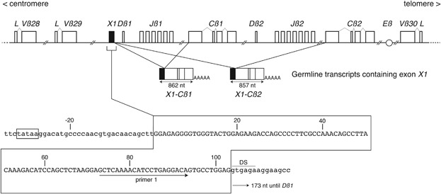 Fig. 1 Exon X1 within the human TCRB locus.1
Key structural properties of TCRB:
- Immunoglobulin folding domain composed of variable region (V), constant region (C) and linker region
- Across the membrane zone positive alkaline residue and CD3 complex form stable salt bridge
- Complementary decision area (CDR3) antigen recognition by V (D) J rearrangement of diversity
Functions of TCRB
The TCRB gene encodes the T-cell receptor β chain, and its core function is to mediate adaptive immune responses. This gene generates a diverse receptor library through V(D)J rearrangement, achieving specific recognition of antigens. Its main functions include:
| Function | Description |
| Antigen recognition | The immune response is initiated by specifically binding to the MHC-peptide complex through the complementary determinant (CDR). |
| Signal transduction | Coupled with the CD3 complex, it transmits antigen recognition signals into the interior of T cells. |
| Immune memory | Participate in the formation of the memory T cell pool and provide long-term immune protection. |
| Self-tolerance | Prevent autoimmune diseases by selectively eliminating autoreactive T cells through the thymus. |
| Tumor monitoring | Identify tumor-specific antigens and initiate anti-tumor immune responses. |
The rearrangement efficiency of the TCRB gene directly affects the diversity of the T cell library, and the conservation of its recombinant signal sequence (RSS) ensures the accuracy of the rearrangement process. The abnormality of this gene's function is closely related to autoimmune diseases, immune deficiencies and tumor occurrence.
Applications of TCRB and TCRB Antibody in Literature
1. Sandberg, Yorick, et al. "Lack of common TCRA and TCRB clonotypes in CD8+/TCRαβ+ T-cell large granular lymphocyte leukemia: a review on the role of antigenic selection in the immunopathogenesis of CD8+ T-LGL." Blood cancer journal 4.1 (2014): e172-e172. https://doi.org/10.1038/bcj.2013.70
This study analyzed 26 patients with CD8+/TCRαβ+ T-LGL leukemia. Through the depth calculation of the TCRB CDR3 region, it was found that their clone sequences lacked homology, which formed a sharp contrast with the CD4+ and TCRγδ+ subtypes. This indicates that CD8+/TCRαβ+ T-LGL leukemia may be driven by multiple different antigens, and the etiology is highly heterogeneous.
2. Triaille, Clement, et al. "Paired rheumatoid arthritis synovial biopsies from small and large joints show similar global transcriptomic patterns with enrichment of private specificity TCRB and TCR signaling pathways." Frontiers in immunology 11 (2020): 593083. https://doi.org/10.3389/fimmu.2020.593083
This study analyzed the paired synovial tissue of patients with rheumatoid arthritis. It was found that the TCRB clone types were highly shared between the large and small joints of the same patient, but the degree of T cell infiltration and clone distribution varied significantly among different patients. This suggests that RA has different synovial pathological types, corresponding respectively to systemic antigen-driven and local antigen-driven T cell activation patterns.
3. Lethé, Bernard, et al. "A new transcript in the TCRB locus unveils the human ortholog of the mouse pre‐Dβ1 promoter." Immunity, Inflammation and Disease 5.3 (2017): 346-354. https://doi.org/10.1002/iid3.172
In this study, a novel non-coding germline transcript composed of a new exon X1 and the Cβ gene was discovered in the human TCRB locus. This transcript is expressed before the V-DJ rearrangement of TCRB is completed. Its promoter may promote the rearrangement process of TCRB by enhancing the accessibility of the Dβ1 region.
4. Gao, Shouguo, et al. "Single-cell RNA sequencing coupled to TCR profiling of large granular lymphocyte leukemia T cells." Nature Communications 13.1 (2022): 1982. https://doi.org/10.1038/s41467-022-29175-x
This study analyzed patients with T-LGLL through single-cell sequencing. The results showed that the diversity of TCRB in patients was significantly reduced, but no shared clone types were found. Clonal expansion is closely related to the down-regulation of the apoptotic pathway. Although Alemtuzumab treatment can upregulate apoptotic genes, it does not eliminate the clonality of TCRB. Its baseline expression can predict the therapeutic effect.
5. Konstantinovsky, Thomas, and Gur Yaari. "A novel approach to T-cell receptor beta chain (TCRB) repertoire encoding using lossless string compression." Bioinformatics 39.7 (2023): btad426. https://doi.org/10.1093/bioinformatics/btad426
This study proposes a new method for TCRB lineage analysis based on the LZ76 algorithm. This method can construct graph models, identify feature sequences, and achieve unified encoding of individual TCRB lineages, providing a new tool for applications such as diversity assessment and sequence generation.
Creative Biolabs: TCRB Antibodies for Research
Creative Biolabs specializes in the production of high-quality TCRB antibodies for research and industrial applications. Our portfolio includes monoclonal antibodies tailored for ELISA, Flow Cytometry, Western blot, immunohistochemistry, and other diagnostic methodologies.
- Custom TCRB Antibody Development: Tailor-made solutions to meet specific research requirements.
- Bulk Production: Large-scale antibody manufacturing for industry partners.
- Technical Support: Expert consultation for protocol optimization and troubleshooting.
- Aliquoting Services: Conveniently sized aliquots for long-term storage and consistent experimental outcomes.
For more details on our TCRB antibodies, custom preparations, or technical support, contact us at email.
Reference
- Lethé, Bernard, et al. "A new transcript in the TCRB locus unveils the human ortholog of the mouse pre‐Dβ1 promoter." Immunity, Inflammation and Disease 5.3 (2017): 346-354. https://doi.org/10.1002/iid3.172
Anti-TCRB antibodies
 Loading...
 Compare




Hot products 
- AActivation
- AGAgonist
- APApoptosis
- BBlocking
- BABioassay
- BIBioimaging
- CImmunohistochemistry-Frozen Sections
- CIChromatin Immunoprecipitation
- CTCytotoxicity
- CSCostimulation
- DDepletion
- DBDot Blot
- EELISA
- ECELISA(Cap)
- EDELISA(Det)
- ESELISpot
- EMElectron Microscopy
- FFlow Cytometry
- FNFunction Assay
- GSGel Supershift
- IInhibition
- IAEnzyme Immunoassay
- ICImmunocytochemistry
- IDImmunodiffusion
- IEImmunoelectrophoresis
- IFImmunofluorescence
- IGImmunochromatography
- IHImmunohistochemistry
- IMImmunomicroscopy
- IOImmunoassay
- IPImmunoprecipitation
- ISIntracellular Staining for Flow Cytometry
- LALuminex Assay
- LFLateral Flow Immunoassay
- MMicroarray
- MCMass Cytometry/CyTOF
- MDMeDIP
- MSElectrophoretic Mobility Shift Assay
- NNeutralization
- PImmunohistologyp-Paraffin Sections
- PAPeptide Array
- PEPeptide ELISA
- PLProximity Ligation Assay
- RRadioimmunoassay
- SStimulation
- SESandwich ELISA
- SHIn situ hybridization
- TCTissue Culture
- WBWestern Blot
