
CLEC7A Antibodies
Background
The CLEC7A gene encodes a pattern recognition receptor of the C-type lectin family, which is mainly expressed on the surface of myeloid immune cells. This receptor can specifically recognize the β-glucan structure in the fungal cell wall and, by activating the spleen tyrosine kinase signaling pathway, trigger downstream inflammatory responses and phagocytosis, thereby initiating the body's innate immune defense against fungal infections. In 2001, researchers first cloned and identified the human CLEC7A gene and revealed its crucial role in anti-fungal immunity. Subsequent studies further confirmed that the deficiency of this gene would increase the susceptibility to chronic skin and mucosal candidiasis. Moreover, CLEC7A can also recognize certain non-classical ligands on the surface of some tumor cells. In recent years, its regulatory function in connecting innate immunity and anti-tumor responses has attracted much attention, providing new molecular targets for the immunotherapy of infectious diseases and cancer.
Structure of CLEC7A
The protein encoded by the CLEC7A gene has a molecular weight of approximately 28 kDa, and there are certain differences among different species. This protein mainly functions as a pattern recognition receptor and initiates the immune response by recognizing components of fungal cell walls.
| Species | Human | Mouse | Rat | Pig | Monkey |
| Molecular Weight (kDa) | 28 | 27 | 27 | 28 | 28 |
| Primary Structural Differences | Extracellular C-type lectin-like domain | Internal ITAM-like motif | Tightly conserved transmembrane region | Consistent key sites for carbohydrate recognition | High homology to humans |
The CLEC7A protein consists of 247 amino acids. Its extracellular segment forms a compact spherical conformation through C-type lectin folding. This protein contains a carbohydrate recognition domain, which is stabilized by calcium ions for ligand binding conformation. The intracellular segment of the receptor has an ITAM-like motif, which can recruit the Syk kinase to initiate signal transduction. The hydrophobic residues in the transmembrane region ensure the correct localization of the receptor on the membrane, while the membrane proximal stem loop region promotes receptor oligomerization and enhances the binding affinity with fungal ligands.
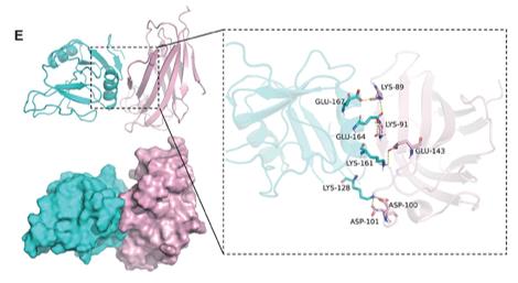 Fig. 1 The interaction between Clec7a (cyan) and MD2 (pink).1
Key structural properties of CLEC7A:
- Extracellular C-type lectin-like domain
- Calcium ion-dependent ligand binding site
- Hydrophobic anchor sequence in the transmembrane region
- Intracellular ITAM-like motif mediating signal transduction
Functions of CLEC7A
The main function of CLEC7A is to recognize the components of fungal cell walls and trigger an immune response. Additionally, it is involved in regulating inflammatory responses and the activation of adaptive immunity.
| Function | Description |
| Pattern Recognition | CLEC7A specifically binds to fungal β-glucan and recognizes invading pathogens. |
| Signal Transduction | Activates the downstream pathway of Syk kinase, inducing the production of inflammatory factors. |
| Phagocytosis | Promotes the uptake and clearance of fungal particles by immune cells. |
| Immune regulation | Inhibits the differentiation of Th17 cells and enhances acquired immunity against fungi. |
| Tumor Monitoring | Identify tumor-associated ligands and participate in anti-tumor immune responses. |
The ligand binding curve of CLEC7A exhibits typical saturation characteristics, which is different from the nonlinear activation pattern of immune receptors. This reflects its high affinity and rapid response characteristics as a pattern recognition receptor for fungal β-glucans.
Applications of CLEC7A and CLEC7A Antibody in Literature
1. Wan, Hanxi, et al. "Clec7a Worsens Long‐Term Outcomes after Ischemic Stroke by Aggravating Microglia‐Mediated Synapse Elimination." Advanced science 11.36 (2024): 2403064. https://doi.org/10.1002/advs.202403064
This study reveals that after ischemic stroke, the expression of CLEC7A in microglia is upregulated, enabling it to phagocytose excitatory synapses. Regulating CLEC7A can reduce synaptic loss and improve neurological function. CLEC7A may interact with the neuronal MD2, making it a potential therapeutic target for IS.
2. Ma, Jie, et al. "Clec7a drives gut fungus-mediated host lipid deposition." Microbiome 11.1 (2023): 264. https://doi.org/10.1186/s40168-023-01698-5
The research reveals that intestinal fungi regulate obesity through the CLEC7A signaling pathway. Supplementing mice with specific fungi can prevent obesity, while activating CLEC7A accelerates obesity, and knocking out this gene can resist obesity. The fungal-CLEC7A axis is a potential target for metabolic regulation.
3. Wang, Yaqiong, et al. "Clec7a expression in inflammatory macrophages orchestrates progression of acute kidney injury." Frontiers in immunology 13 (2022): 1008727. https://doi.org/10.3389/fimmu.2022.1008727
The study reveals that macrophages after renal IRI can be classified into four subgroups. Clec7a+ M1 cells have strong pro-inflammatory phagocytic ability, while Clec7a- M2 cells promote repair. Regulating the expression of Clec7a can improve renal function, suggesting that it is a new target for the treatment of AKI.
4. Yang, Shubing, et al. "Clec7a signaling in microglia promotes synapse loss associated with tauopathy." International Journal of Molecular Sciences 26.7 (2025): 2888. https://doi.org/10.3390/ijms26072888
The study found that the Clec7a-SYK signaling axis was activated in the tau protein disease model, and it mediated the clearance of synapses by microglia. Inhibiting Clec7a could alleviate neuroinflammation, restore synaptic function and improve memory. This suggests that Clec7a is a potential therapeutic target for synaptic damage associated with tau protein diseases.
5. Wang, Jinchao, et al. "CLEC7A regulates M2 macrophages to suppress the immune microenvironment and implies poorer prognosis of glioma." Frontiers in immunology 15 (2024): 1361351. https://doi.org/10.3389/fimmu.2024.1361351
The study found that CLEC7A is highly expressed in gliomas and is positively correlated with malignancy, serving as an independent prognostic factor. It affects the immune microenvironment by regulating the chemotaxis and polarization of M2-type macrophages, and is expected to become a new target for immunotherapy in gliomas.
Creative Biolabs: CLEC7A Antibodies for Research
Creative Biolabs specializes in the production of high-quality CLEC7A antibodies for research and industrial applications. Our portfolio includes monoclonal and polyclonal antibodies tailored for ELISA, Flow Cytometry, Western blot, immunohistochemistry, and other diagnostic methodologies.
- Custom CLEC7A Antibody Development: Tailor-made solutions to meet specific research requirements.
- Bulk Production: Large-scale antibody manufacturing for industry partners.
- Technical Support: Expert consultation for protocol optimization and troubleshooting.
- Aliquoting Services: Conveniently sized aliquots for long-term storage and consistent experimental outcomes.
For more details on our CLEC7A antibodies, custom preparations, or technical support, contact us at email.
Reference
- Wan, Hanxi, et al. "Clec7a Worsens Long‐Term Outcomes after Ischemic Stroke by Aggravating Microglia‐Mediated Synapse Elimination." Advanced science 11.36 (2024): 2403064. Distributed under Open Access license CC BY 4.0. Cropped from the original figure. https://doi.org/10.1002/advs.202403064
Anti-CLEC7A antibodies
 Loading...
 Compare




Hot products 
- AActivation
- AGAgonist
- APApoptosis
- BBlocking
- BABioassay
- BIBioimaging
- CImmunohistochemistry-Frozen Sections
- CIChromatin Immunoprecipitation
- CTCytotoxicity
- CSCostimulation
- DDepletion
- DBDot Blot
- EELISA
- ECELISA(Cap)
- EDELISA(Det)
- ESELISpot
- EMElectron Microscopy
- FFlow Cytometry
- FNFunction Assay
- GSGel Supershift
- IInhibition
- IAEnzyme Immunoassay
- ICImmunocytochemistry
- IDImmunodiffusion
- IEImmunoelectrophoresis
- IFImmunofluorescence
- IGImmunochromatography
- IHImmunohistochemistry
- IMImmunomicroscopy
- IOImmunoassay
- IPImmunoprecipitation
- ISIntracellular Staining for Flow Cytometry
- LALuminex Assay
- LFLateral Flow Immunoassay
- MMicroarray
- MCMass Cytometry/CyTOF
- MDMeDIP
- MSElectrophoretic Mobility Shift Assay
- NNeutralization
- PImmunohistologyp-Paraffin Sections
- PAPeptide Array
- PEPeptide ELISA
- PLProximity Ligation Assay
- RRadioimmunoassay
- SStimulation
- SESandwich ELISA
- SHIn situ hybridization
- TCTissue Culture
- WBWestern Blot
