
ID1 Antibodies
Background
The ID1 gene encodes a protein belonging to the helical-ring-helix transcription factor family and is widely present in various human tissues. This protein regulates gene expression by forming heterodimers with other transcription factors, thereby influencing key life processes such as cell differentiation, proliferation and apoptosis. During embryonic development, the ID1 gene plays a significant regulatory role in neural tissue and blood vessel formation, and its abnormal expression is closely related to the occurrence and development of various malignant tumors. This gene was first identified by Robert Benezra's team in 1990. Its naming abbreviation "ID" is derived from its functional characteristic - the ability to inhibit the process of cell differentiation. As a key node in the cell signal transduction pathway, the ID1 gene has become an important model system in tumor biology and developmental biology research, providing an important perspective for revealing the gene expression regulatory network and its mechanism of action in diseases.
Structure of ID1
The ID1 gene encodes a protein with a molecular weight of approximately 14-16 kDa, and its precise molecular weight varies depending on post-translational modifications and experimental conditions. This protein belongs to the family of helical-ring-helix transcription factors, and its most notable structural feature is the lack of a DNA-binding domain.
| Species | Human | Mouse | Rat | Zebrafish | African clawed toad |
| Molecular Weight (kDa) | 15.8 | 15.6 | 15.7 | 16.2 | 15.9 |
| Primary Structural Differences | Typical structure containing 154 amino acids | N-terminal sequence has three amino acid differences | Phosphorylation site variation exists in the C-terminal region | Dimerization domains have relatively high conservation | There are adaptive mutations in the helical region |
The ID1 protein is composed of approximately 154 amino acids and exhibits a typical helical-ring-helical folded conformation. Its three-dimensional structure consists of two α -helices connected by a ring region, forming a unique dimerization interface. The core function of this protein lies in its unique design lacking an alkaline DNA binding region. This structural feature enables it to form a heterodimer with the bHLH transcription factor, which has DNA binding ability, through a dominant negative regulatory mechanism, thereby inhibiting the transcriptional activity of the target gene. This structural design serves as the functional basis for the ID1 protein's participation in cell cycle regulation and differentiation inhibition.
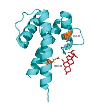 Fig. 1 Predicted UDCA-ID1 interaction.1
Key structural properties of ID1:
- Helix-loop-helix (HLH) dimerization domain
- Lack of DNA-binding regions (basic domains)
- Conserved protein-protein interaction interface
Functions of ID1
The main function of the ID1 protein is to serve as a key negative regulatory factor for cell differentiation. However, it is also widely involved in various physiological and pathological processes, including cell cycle regulation, tumorigenesis and angiogenesis.
| Function | Description |
| Differentiation inhibition | By forming heterodimers with other bHLH transcription factors, it inhibits the expression of tissue-specific genes and prevents terminal cell differentiation. |
| Proliferation promotion | Remove the expression restrictions of differentiation-related genes, promote cells to continuously enter the cell cycle, and enhance proliferation capacity. |
| Regulation of embryonic development | High expression in early embryonic development, maintaining a variety of precursor cells undifferentiated state, instruction organization form. |
| Tumorigenesis participation | In a variety of abnormally high expression in cancer, inhibit differentiation, promote the proliferation and angiogenesis, drive tumor progression. |
| Angiogenesis support | In response to growth factor signals, it upregulates pro-angiogenic factors and promotes angiogenesis in embryos and tumor tissues. |
The functional realization of the ID1 protein does not rely on direct DNA binding, but rather through its unique "dominant negative regulation" mechanism - that is, by binding to other transcription factors with DNA binding capabilities, it forms non-functional dimers that cannot effectively activate target genes, thereby achieving precise control over the cell differentiation program. This regulatory mechanism makes it hold a core position in developmental biology and tumor biology.
Applications of ID1 and ID1 Antibody in Literature
1. Lee, Woo Ho, et al. "Transcription factor Id1 plays an essential role in Th9 cell differentiation by inhibiting Tcf3 and Tcf4." Advanced Science 10.35 (2023): 2305527. https://doi.org/10.1002/advs.202305527
The article indicates that ID1 is a key transcription factor in the differentiation of Th9 cells. It is induced by IL-4 and TGF-β, and promotes the expression of interleukin-9 by antagonizing the inhibitory effect of the transcriptional suppressor Tcf3/Tcf4 on the Il9 gene, thereby driving the differentiation of Th9 cells and affecting asthma inflammation.
2. Zhao, Zhengxiao, et al. "Inhibitor of differentiation 1 (Id1) in cancer and cancer therapy." International journal of medical sciences 17.8 (2020): 995. https://doi.org/10.7150/ijms.42805
The article indicates that ID1, as a stem cell-like gene, is highly expressed in various cancers. It significantly promotes tumor proliferation, angiogenesis, metastasis and drug resistance by activating multiple signaling pathways such as EGFR and PI3K/Akt, and is thus regarded as a highly promising new target for anti-tumor treatment.
3. Kantzer, Christina Geraldine, et al. "ID1 and CEBPA coordinate epidermal progenitor cell differentiation." Development 149.22 (2022): dev201262. https://doi.org/10.1242/dev.201262
The article indicates that ID1 is a key factor coordinating epidermal development and is enriched in basal progenitor cells. It functions by inhibiting differentiation, maintaining the proliferation of progenitor cells and the adhesion of the basement membrane. ID1 interacts with bHLH transcription factors (such as TCF3) and antagonize with the differentiation mediator CEBPA, jointly balancing the proliferation and differentiation process of progenitor cells.
4. Phadte, Pratham, et al. "Autophagy-mediated ID1 turnover dictates chemo-resistant fate in ovarian cancer stem cells." Journal of Experimental & Clinical Cancer Research 43.1 (2024): 222. https://doi.org/10.1186/s13046-024-03147-z
The article indicates that in ovarian cancer, autophagy can degrade the stem cell regulatory factor ID1, driving drug-resistant stem cells to differentiate into sensitive cells. After ID1 degrades, it releases the transcription factor TCF12 it binds to, which in turn activates the expression of the cisplatin drug inflow transporter SLC31A1, increases intracellular drug accumulation, and ultimately reverses chemotherapy resistance.
5. Meng, Jiao, et al. "ID1 confers cancer cell chemoresistance through STAT3/ATF6-mediated induction of autophagy." Cell death & disease 11.2 (2020): 137. https://doi.org/10.1038/s41419-020-2327-1
The article indicates that in ovarian cancer, ID1 activates the NF-κB/IL-6 axis, thereby driving STAT3 phosphorylation and upregulating ATF6 transcription. This signaling axis induces endoplasmic reticulum stress and autophagy, ultimately leading to cancer cells developing resistance to cisplatin and paclitaxel. Moreover, high expression of ID1 or ATF6 is significantly associated with poor prognosis in patients.
Creative Biolabs: ID1 Antibodies for Research
Creative Biolabs specializes in the production of high-quality ID1 antibodies for research and industrial applications. Our portfolio includes monoclonal antibodies tailored for ELISA, Flow Cytometry, Western blot, immunohistochemistry, and other diagnostic methodologies.
- Custom ID1 Antibody Development: Tailor-made solutions to meet specific research requirements.
- Bulk Production: Large-scale antibody manufacturing for industry partners.
- Technical Support: Expert consultation for protocol optimization and troubleshooting.
- Aliquoting Services: Conveniently sized aliquots for long-term storage and consistent experimental outcomes.
For more details on our ID1 antibodies, custom preparations, or technical support, contact us at email.
Reference
- Dong, Xi, et al. "Ursodesoxycholic acid alleviates liver fibrosis via proregeneration by activation of the ID1‐WNT2/HGF signaling pathway." Clinical and Translational Medicine 11.2 (2021): e296. https://doi.org/10.1002/ctm2.296
Anti-ID1 antibodies
 Loading...
 Compare




Hot products 
- AActivation
- AGAgonist
- APApoptosis
- BBlocking
- BABioassay
- BIBioimaging
- CImmunohistochemistry-Frozen Sections
- CIChromatin Immunoprecipitation
- CTCytotoxicity
- CSCostimulation
- DDepletion
- DBDot Blot
- EELISA
- ECELISA(Cap)
- EDELISA(Det)
- ESELISpot
- EMElectron Microscopy
- FFlow Cytometry
- FNFunction Assay
- GSGel Supershift
- IInhibition
- IAEnzyme Immunoassay
- ICImmunocytochemistry
- IDImmunodiffusion
- IEImmunoelectrophoresis
- IFImmunofluorescence
- IGImmunochromatography
- IHImmunohistochemistry
- IMImmunomicroscopy
- IOImmunoassay
- IPImmunoprecipitation
- ISIntracellular Staining for Flow Cytometry
- LALuminex Assay
- LFLateral Flow Immunoassay
- MMicroarray
- MCMass Cytometry/CyTOF
- MDMeDIP
- MSElectrophoretic Mobility Shift Assay
- NNeutralization
- PImmunohistologyp-Paraffin Sections
- PAPeptide Array
- PEPeptide ELISA
- PLProximity Ligation Assay
- RRadioimmunoassay
- SStimulation
- SESandwich ELISA
- SHIn situ hybridization
- TCTissue Culture
- WBWestern Blot
