IL18 Antibodies
Background
IL18 gene encoding a called interleukin - 18 proinflammatory cytokines, the gene is mainly expressed in active immune cells and epithelial cells. Its products, by binding to specific receptors, can activate the NF-κB signaling pathway, thereby inducing the production of cytokines such as interferon - γ, playing a core role in the host's resistance to pathogen infection and immune regulation. This gene was officially identified in 1996 and is an important member of the interleukin-1 superfamily. Subsequent studies have found that it not only participates in the bridging role between innate immunity and acquired immunity, but also plays a dual role in pathological processes such as autoimmune diseases, metabolic syndrome and tumor microenvironment regulation. Therefore, it has become an important research object for therapeutic targets of various immune-related diseases.
Structure of IL18
The protein encoded by the IL18 gene is a pro-inflammatory cytokine with a molecular weight of approximately 22.2 kDa. There are certain differences in this molecular weight among different species, mainly due to the processing and maturation process of its precursor protein and the subtle differences in amino acid sequences.
| Species | Human | Mouse | Rat |
| Molecular Weight (kDa) | 22.2 | 22.3 | 22.1 |
| Primary Structural Differences | Mature body of 157 amino acids, active form of homologous dimers | With the human IL - 18 about 65% sequence homology, IL - 18 r can be combined with people | Similar to human functions, it is often used in disease model research |
The primary structure of this protein folds to form a typical β -clover stereoconfiguration. Its spatial structure is composed of 12 β -folded sheets forming the core skeleton, which are connected through multiple annular regions. Specific regions on the protein surface are responsible for binding to the IL-18 receptor α chain, and the conformational accuracy of this binding site is crucial for initiating downstream signal transduction. A key structural functional site is located between Asp53 and Lys89, which stabilize the receptor-binding interface by forming a salt bridge, thereby regulating its powerful inflammation-inducing ability.
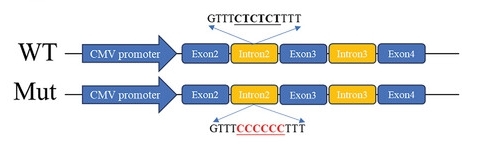 Fig. 1 Schematic diagram of the IL-18 minigene constructs.1
Key structural properties of IL18:
- Typical β-clover topology
- The core framework is composed of 12 β -folded sheets
- Surface receptor binding interface determines its signal specificity
Functions of IL18
The core function of the protein encoded by the IL18 gene is to activate immune responses and inflammatory reactions. However, this factor also plays a dual role in various pathological processes, including anti-infection, anti-tumor, and participation in the occurrence and development of autoimmune diseases.
| Function | Description |
| Immune activation | Induce T cells and NK cells to produce interferon - γ, enhance cellular immune responses, and combat intracellular pathogens. |
| Inflammatory trigger | As a pro-inflammatory factor, it activates the NF-κB signaling pathway and promotes the release of inflammatory mediators such as TNF-α and IL-1. |
| Anti-tumor effect | By enhancing the cytotoxicity of effector immune cells, tumor growth is inhibited, but its specific role in the tumor microenvironment is bidirectional. |
| Metabolic regulation | Participate in obesity and related metabolic inflammation process, affect the insulin sensitivity. |
| Tissue repair | In the later stage of inflammation, it indirectly affects the process of tissue regeneration and fibrosis by regulating other growth factors. |
Unlike most interleukins, IL18 is stored in the cytoplasm as an inactive precursor and needs to be cleaved by proteases such as caspase-1 to mature and activate. This characteristic makes it a precisely regulated key node in the inflammatory response.
Applications of IL18 and IL18 Antibody in Literature
1. Zhao, Cheng, et al. "PTBP3 Mediates IL‐18 Exon Skipping to Promote Immune Escape in Gallbladder Cancer." Advanced Science 11.38 (2024): 2406633. https://doi.org/10.1002/advs.202406633
The article indicates that in gallbladder cancer, PTBP3 mediates the skipping of IL-18 exon to produce the oncogenic isomer ΔIL-18, which promotes PD-1 stability by down-regulating FBXO38 in CD8+T cells, leading to immune escape. Targeting ΔIL-18 can inhibit tumor growth and provide new ideas for immunotherapy.
2. Choi, Yeon Ho, et al. "IL-27 enhances IL-15/IL-18-mediated activation of human natural killer cells." Journal for immunotherapy of cancer 7.1 (2019): 168. https://doi.org/10.1186/s40425-019-0652-7
This study found that when human NK cells were expanded in vitro, the combination of IL-18, IL-15 and IL-27 had a significant synergistic effect, which could most effectively enhance the proliferation, cytotoxicity and IFN-γ secretion of NK cells, and increase the expression of their activated receptors, providing an optimized culture protocol for cancer immunotherapy.
3. Berjis, Abdulla, et al. "Pretreatment with IL-15 and IL-18 rescues natural killer cells from granzyme B-mediated apoptosis after cryopreservation." Nature communications 15.1 (2024): 3937. https://doi.org/10.1038/s41467-024-47574-0
Research has found that before cryopreserving NK cells, combined pretreatment with IL-18 and IL-15 can effectively inhibit apoptosis by inducing anti-apoptotic genes and temporarily reducing the level of granzyme B, increase the cell recovery rate to over 90%, and maintain their anti-tumor activity.
4. Rahman, Ayesha, and Lingadahalli S. Shashidhara. "Analyzing the influence of IL18 in regulation of YAP1 in breast oncogenesis using cBioportal." Cancer Reports 5.3 (2022): e1484. https://doi.org/10.1002/cnr2.1484
The article indicates that research based on TCGA data has found that in breast cancer, IL-18 participates in tumor progression by influencing the expression of YAP1. High expression of IL-18 predicts a better prognosis, while high YAP1 accompanied by low IL-18 leads to adverse consequences, suggesting that IL-18 may serve as a potential therapeutic target for YAP1-overexpressing breast cancer.
5. Sato, Paula Keiko, et al. "Polymorphism in the Promoter Region of the IL18 Gene and the Association with Severity on Paracoccidioidomycosis." Frontiers in immunology 11 (2020): 542210. https://doi.org/10.3389/fimmu.2020.542210
This study, through the analysis of patients with coccidiosis, found that the A allele (rs1946518) at the -607 locus of the IL-18 gene significantly increases the risk of acute and multifocal chronic types, while the C allele may play a protective role against the single-focal chronic type, revealing the important role of this gene polymorphism in the clinical outcomes of the disease.
Creative Biolabs: IL18 Antibodies for Research
Creative Biolabs specializes in the production of high-quality IL18 antibodies for research and industrial applications. Our portfolio includes monoclonal antibodies tailored for ELISA, Flow Cytometry, Western blot, immunohistochemistry, and other diagnostic methodologies.
- Custom IL18 Antibody Development: Tailor-made solutions to meet specific research requirements.
- Bulk Production: Large-scale antibody manufacturing for industry partners.
- Technical Support: Expert consultation for protocol optimization and troubleshooting.
- Aliquoting Services: Conveniently sized aliquots for long-term storage and consistent experimental outcomes.
For more details on our IL18 antibodies, custom preparations, or technical support, contact us at email.
Reference
- Zhao, Cheng, et al. "PTBP3 Mediates IL‐18 Exon Skipping to Promote Immune Escape in Gallbladder Cancer." Advanced Science 11.38 (2024): 2406633. https://doi.org/10.1002/advs.202406633
Anti-IL18 antibodies
 Loading...


Hot products 
-
Rabbit Anti-BRCA2 Recombinant Antibody (D9S6V) (CBMAB-CP0017-LY)

-
Mouse Anti-dsRNA Recombinant Antibody (2) (CBMAB-D1807-YC)

-
Mouse Anti-ACLY Recombinant Antibody (V2-179314) (CBMAB-A0610-YC)

-
Mouse Anti-CCDC6 Recombinant Antibody (CBXC-0106) (CBMAB-C5397-CQ)

-
Mouse Anti-CTNND1 Recombinant Antibody (CBFYC-2414) (CBMAB-C2487-FY)

-
Rat Anti-CD63 Recombinant Antibody (7G4.2E8) (CBMAB-C8725-LY)

-
Mouse Anti-G6PD Recombinant Antibody (13B331) (CBMAB-G1553-LY)

-
Mouse Anti-AP4E1 Recombinant Antibody (32) (CBMAB-A2996-YC)

-
Rat Anti-CD300A Recombinant Antibody (172224) (CBMAB-C0423-LY)

-
Mouse Anti-ASH1L Monoclonal Antibody (ASH5H03) (CBMAB-1372-YC)

-
Mouse Anti-BIRC3 Recombinant Antibody (16E63) (CBMAB-C3367-LY)

-
Rabbit Anti-CCL5 Recombinant Antibody (R0437) (CBMAB-R0437-CN)

-
Mouse Anti-ATP1A2 Recombinant Antibody (M7-PB-E9) (CBMAB-A4013-YC)

-
Mouse Anti-8-oxoguanine Recombinant Antibody (V2-7719) (CBMAB-1898CQ)

-
Mouse Anti-DLL4 Recombinant Antibody (D1090) (CBMAB-D1090-YC)

-
Rabbit Anti-B2M Recombinant Antibody (CBYY-0059) (CBMAB-0059-YY)

-
Rabbit Anti-ABL1 (Phosphorylated Y185) Recombinant Antibody (V2-443434) (PTM-CBMAB-0001YC)

-
Mouse Anti-CIITA Recombinant Antibody (CBLC160-LY) (CBMAB-C10987-LY)

-
Mouse Anti-ALB Recombinant Antibody (V2-180650) (CBMAB-A2186-YC)

-
Mouse Anti-ARID1B Recombinant Antibody (KMN1) (CBMAB-A3546-YC)

- AActivation
- AGAgonist
- APApoptosis
- BBlocking
- BABioassay
- BIBioimaging
- CImmunohistochemistry-Frozen Sections
- CIChromatin Immunoprecipitation
- CTCytotoxicity
- CSCostimulation
- DDepletion
- DBDot Blot
- EELISA
- ECELISA(Cap)
- EDELISA(Det)
- ESELISpot
- EMElectron Microscopy
- FFlow Cytometry
- FNFunction Assay
- GSGel Supershift
- IInhibition
- IAEnzyme Immunoassay
- ICImmunocytochemistry
- IDImmunodiffusion
- IEImmunoelectrophoresis
- IFImmunofluorescence
- IGImmunochromatography
- IHImmunohistochemistry
- IMImmunomicroscopy
- IOImmunoassay
- IPImmunoprecipitation
- ISIntracellular Staining for Flow Cytometry
- LALuminex Assay
- LFLateral Flow Immunoassay
- MMicroarray
- MCMass Cytometry/CyTOF
- MDMeDIP
- MSElectrophoretic Mobility Shift Assay
- NNeutralization
- PImmunohistologyp-Paraffin Sections
- PAPeptide Array
- PEPeptide ELISA
- PLProximity Ligation Assay
- RRadioimmunoassay
- SStimulation
- SESandwich ELISA
- SHIn situ hybridization
- TCTissue Culture
- WBWestern Blot
