
SMC3 Antibodies
Background
SMC3 is an important member of the chromosome structure maintenance protein (SMC) family and mainly exists in the nucleus of eukaryotes. As a core component of the adhesive protein complex, it forms a circular structure together with SMC1, RAD21 and STAG proteins, responsible for the adhesion of sister chromatids and ensuring the precise distribution of chromosomes during cell division. Research has found that the acetylation modification of SMC3 can dynamically regulate the higher-order structure of chromatin, influencing gene expression and DNA damage repair. The abnormal function of this protein is closely related to various developmental disorders (such as Cornelia de Lange syndrome) and cancer. As a key molecule for maintaining genomic stability, SMC3 provides an important research model for understanding the dynamic regulation of chromosomes and the mechanisms of cell division.
Structure of SMC3
SMC3 is a large chromosomal structure maintenance protein with a molecular weight of approximately 140 kDa, and its molecular weight is highly conserved across different species. The following table shows the comparison of key features of SMC3 in different species:
| Species | Human | Mice | Fruit flies | yeast |
| Molecular Weight (kDa) | 141.5 | 140.8 | 139.2 | 138.6 |
| Primary Structural Differences | Conserved ATPase domain | Highly similar coiled spiral zones | Core functional areas remain | Simplified but fully functional |
The SMC3 protein is composed of approximately 1,200 amino acids and features a typical SMC protein structure: there is an ATPase domain at both the N-terminal and C-terminal, which are connected by a long coily-helical region in the middle, forming an antiparallel dimer. The core function of this protein depends on its unique V-shaped conformation, which is stabilized by intramolecular disulfide bonds. The ATPase domain of SMC3 contains Walker A and Walker B mosomes, which are responsible for the energy-dependent chromatin loop extrusion process. The hinge domain in its coiled helical region is crucial for the dynamic conformational changes of the protein, enabling SMC3 to participate in the advanced structural regulation of chromatin.
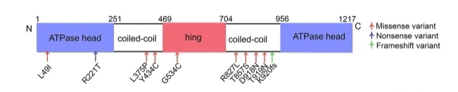 Fig. 1 Protein domain plot of SMC3 and the site (top panel).1
Key structural properties of SMC3:
- Characteristic V-shaped dimer structure
- Conserved ATP binding sites
- Intramolecular disulfide bonds
- Hinge domain
- Acetylation modification site (Lys105/106)
Functions of SMC3
The core function of the SMC3 protein is to maintain chromosome structure and regulate gene expression. Its main functions are shown in the following table:
| Function | Description |
| Sister chromatids are adhered | Ensure the correct pairing and separation of sister chromatids during mitosis and meiosis. |
| Chromatin ring formation | Mediated by cohesin complexes chromatin ring out, control gene three-dimensional space organization. |
| DNA damage repair | Participate in homologous recombination repair and maintain genomic stability. |
| Regulation of gene expression | Influence the interaction between transcription factors and enhancers by altering the spatial conformation of chromatin. |
| Cell cycle regulation | Interacts with kinases such as CDK1 to coordinate mitotic progression. |
The functional exertion of SMC3 depends on its ATPase activity. Unlike typical adhesives, the chromatin ring extrudation mediated by SMC3 shows a continuous rather than stepwise characteristic, which is closely related to its special role in gene regulation. During heart development, SMC3 influences organ formation by regulating the expression of key transcription factors such as TBX5.
Applications of SMC3 and SMC3 Antibody in Literature
1. Ghiselli, Giancarlo. "SMC3 knockdown triggers genomic instability and p53-dependent apoptosis in human and zebrafish cells." Molecular Cancer 5.1 (2006): 52. https://doi.org/10.1186/1476-4598-5-52
The article indicates that the deletion of SMC3 protein induces apoptosis through the p53-Bax pathway, leading to abnormal embryonic development and centrosome amplification, and causing chromosomal instability. Studies on SMC3 antibodies have confirmed that their absence activates p53-dependent mitotic checkpoints and promotes aneuploidy formation.
2. Laugsch, Magdalena, et al. "Imbalance of SMC1 and SMC3 cohesins causes specific and distinct effects." PLoS One 8.6 (2013): e65149. https://doi.org/10.1371/journal.pone.0065149
The article indicates that studies on SMC3 antibodies have found that SMC3 knockout leads to the instability of SMC1 and its failure to accumulate in the cytoplasm, while SMC3 remains in the cytoplasm after SMC1 knockout. The absence of both leads to the degradation of RAD21, but the chromatin binding characteristics are significantly different, suggesting that SMC1/SMC3 has non-redundant functions in the regulation of adhesive proteins.
3. Zhang, Bowen, et al. "SMC3 contributes to heart development by regulating super-enhancer associated genes." Experimental & molecular medicine 56.8 (2024): 1826-1842. https://doi.org/10.1038/s12276-024-01293-0
The article indicates that the research on SMC3 antibodies reveals its crucial role in cardiac development: SMC3 mutations lead to congenital heart disease and affect the expression of genes such as Ets2 by regulating the interaction between super enhancers and promoters. The deletion of SMC3 causes abnormal outflow tract, providing a new mechanism for cardiac development.
4. Ghiselli, Giancarlo, and Chang-Gong Liu. "Global gene expression profiling of cells overexpressing SMC3." Molecular cancer 4.1 (2005): 34. https://doi.org/10.1186/1476-4598-4-34
The article indicates that overexpression of SMC3 promotes tumorigenesis by activating oncogenes such as RhoB/CRE-BPa and upregulates the ras-rho/cAMP signaling pathway. Studies on SMC3 antibodies have revealed that they enhance transcriptional activity and drive cell transformation through SRE/CRE cis elements.
5. Collier, James E., and Kim A. Nasmyth. "DNA passes through cohesin's hinge as well as its Smc3–kleisin interface." Elife 11 (2022): e80310.https://doi.org/10.7554/eLife.80310
The article indicates that the SMC3 antibody study reveals the dual DNA channel mechanism of the adhesion ring: the Smc3/Scc1 interface serves as the DNA exit, while the Smc1/3 hinge region relies on Scc2/3 to regulate DNA entry, which is crucial for the establishment of sister chromatid adhesion.
Creative Biolabs: SMC3 Antibodies for Research
Creative Biolabs specializes in the production of high-quality SMC3 antibodies for research and industrial applications. Our portfolio includes monoclonal antibodies tailored for ELISA, Flow Cytometry, Western blot, immunohistochemistry, and other diagnostic methodologies.
- Custom SMC3 Antibody Development: Tailor-made solutions to meet specific research requirements.
- Bulk Production: Large-scale antibody manufacturing for industry partners.
- Technical Support: Expert consultation for protocol optimization and troubleshooting.
- Aliquoting Services: Conveniently sized aliquots for long-term storage and consistent experimental outcomes.
For more details on our SMC3 antibodies, custom preparations, or technical support, contact us at email.
Reference
- Zhang, Bowen, et al. "SMC3 contributes to heart development by regulating super-enhancer associated genes." Experimental & molecular medicine 56.8 (2024): 1826-1842. https://doi.org/10.1038/s12276-024-01293-0
Anti-SMC3 antibodies
 Loading...
 Compare




Hot products 
- AActivation
- AGAgonist
- APApoptosis
- BBlocking
- BABioassay
- BIBioimaging
- CImmunohistochemistry-Frozen Sections
- CIChromatin Immunoprecipitation
- CTCytotoxicity
- CSCostimulation
- DDepletion
- DBDot Blot
- EELISA
- ECELISA(Cap)
- EDELISA(Det)
- ESELISpot
- EMElectron Microscopy
- FFlow Cytometry
- FNFunction Assay
- GSGel Supershift
- IInhibition
- IAEnzyme Immunoassay
- ICImmunocytochemistry
- IDImmunodiffusion
- IEImmunoelectrophoresis
- IFImmunofluorescence
- IGImmunochromatography
- IHImmunohistochemistry
- IMImmunomicroscopy
- IOImmunoassay
- IPImmunoprecipitation
- ISIntracellular Staining for Flow Cytometry
- LALuminex Assay
- LFLateral Flow Immunoassay
- MMicroarray
- MCMass Cytometry/CyTOF
- MDMeDIP
- MSElectrophoretic Mobility Shift Assay
- NNeutralization
- PImmunohistologyp-Paraffin Sections
- PAPeptide Array
- PEPeptide ELISA
- PLProximity Ligation Assay
- RRadioimmunoassay
- SStimulation
- SESandwich ELISA
- SHIn situ hybridization
- TCTissue Culture
- WBWestern Blot
