
GNAL Antibodies
Background
The GNAL gene encodes the G protein α subunit olfactory type, which is mainly distributed in the central nervous system, especially in the striatum and dopaminergic pathways. This gene product regulates intracellular cAMP synthesis by modulating G protein-coupled receptor signal transduction, thereby influencing neuronal plasticity and signal integration. Its crucial role in the development and functional maintenance of the nervous system is closely related to the pathogenesis of neuropsychiatric disorders such as Parkinson's disease and schizophrenia. This gene was first identified in 1993. Its unique signal regulation pattern provides an important model for understanding the specific functions of G proteins in the nervous system. Related research continues to promote the analysis of the association mechanism between neurotransmitter pathways and diseases.
Structure of GNAL
The G protein α subunit olfactory type encoded by the GNAL gene is a polypeptide with a molecular weight of approximately 44-45 kDa. Its precise molecular weight varies slightly among different species, mainly due to specific amino acid substitutions within the gene coding region.
| Species | Human | Mouse | Rat | Zebrafish | Macaque |
| Molecular Weight (kDa) | 44.8 | 44.6 | 44.7 | 45.2 | 44.8 |
| Primary Structural Differences | Standard reference sequence | Serine variation at position 215 | Associated with rat dopamine signaling features | The C-terminal domain has fish-specific modificationsb | More than 98% homology to human sequence |
This protein is composed of 381 amino acids, and its three-dimensional structure presents a typical Gα three-domain architecture. The GTP-binding domain located in the center regulates the G protein cycle through conformational changes in the Switch I-III region. The serine residue at position 215, as a phosphorylation modification site, directly affects the activity intensity of the cAMP signaling pathway. The α -helical beam formed by the C-terminal domain is responsible for the specific binding to the Gβγ subunit, and this interaction determines the specificity and efficiency of signal transduction.
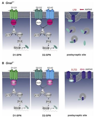 Fig. 1 Schematic representation of Gαolf-mediated biological processes.1
Key structural properties of GNAL:
- Typical folded conformation of the Gα tridomain
- Highly conserved GTP-binding pocket with switch region
- Gβγ subunit binding interface mediated by carboxyl terminal alpha helices
Functions of GNAL
The Gα olf protein encoded by the GNAL gene mainly functions to mediate the signal transduction of olfactory receptors and dopamine receptors, and is also involved in multiple neuroregulatory processes.
| Function | Description |
| Olfactory signal transduction | The ACIII-cAMP pathway is activated in olfactory bulb neurons to convert odor molecular signals into electrical signals. |
| Dopamine signal integration | Enhanced striatal cAMP production via D1/D5 receptors regulates motor coordination and reward behavior. |
| Regulation of neuronal plasticity | After CREB phosphorylation pathways through influence dendritic spines morphogenesis and synaptic transmission efficiency. |
| Maintenance of circadian rhythm | The GPCR signaling network involved in the suprachiasmatic nucleus regulates the oscillations of biological clock gene expression. |
| Association with mental illness | Its functional defect can lead to cAMP signal disorder and is significantly associated with the risk of bipolar disorder. |
The GTP hydrolysis curve of this protein shows a single-phase exponential attenuation feature, which is different from the co-activation mode of most Gα subunits, reflecting its special adaptive mechanism in continuous signal transduction.
Applications of GNAL and GNAL Antibody in Literature
1. Kumar, Ajeet, Samira Saeirad, and Mark S. LeDoux. "Mouse Gnal transcripts and transcriptomics in isolated dystonia." Research Square (2025): rs-3. https://doi.org/10.21203/rs.3.rs-7222154/v1
The article indicates that the two protein subtypes of the GNAL gene have different functions: Gα(olf) dominates olfactory and motor signal transduction, and its mutation has a high risk of causing disease; XLGα(olf) is widely distributed in brain regions and may be related to development, providing a new direction for the study of related diseases.
2. Pelosi, Assunta, et al. "Heterozygous Gnal mice are a novel animal model with which to study dystonia pathophysiology." Journal of Neuroscience 37.26 (2017): 6253-6267. https://doi.org/10.1523/JNEUROSCI.1529-16.2017
The article indicates that insufficient haploid dose of the GNAL gene can lead to dystonia. Research has found that Gnal+/- mice exhibit similar abnormal postures under the action of cholinergic agonists. This effect is mediated by the striatum and can be reversed by M1 receptor antagonists, providing a model for revealing pathological mechanisms.
3. Danquah, Bright D., et al. "Mass Spectrometric analysis of antibody—Epitope peptide complex dissociation: Theoretical concept and practical procedure of binding strength characterization." Molecules 25.20 (2020): 4776. https://doi.org/10.32604/or.2024.045769
The article indicates that low expression of GNAL predicts a poor prognosis for patients with glioma. Its expression level is significantly correlated with the tumor immune microenvironment, DNA methylation and immunotherapy response, and can be used as a potential prognostic biomarker.
4. Charlesworth, Gavin, Kailash P. Bhatia, and Nicholas W. Wood. "No pathogenic GNAL mutations in 192 sporadic and familial cases of cervical dystonia." Movement Disorders 29.1 (2014): 154-155. https://doi.org/10.1002/mds.25713
The article indicates that the new variations identified through GNAL gene screening have not been co-inherited with the disease after family analysis, and thus do not support their status as the cause of the disease. This indicates that the actual incidence of GNAL mutations in dystonia is very low.
5. LONGUEVILLE, Sophie, et al. "Characterization of mice with cell type-specific Gnal loss of function provides insights on GNAL-linked dystonia." bioRxiv (2025): 2025-07. https://doi.org/10.1016/j.nbd.2025.107071
The article indicates that Gαolf regulates motor coordination and spontaneous activity respectively in D1-SPN and A2A-SPN neurons, and its specific absence leads to different behavioral phenotypes, revealing the potential cellular mechanism of GNA-related dystonia.
Creative Biolabs: GNAL Antibodies for Research
Creative Biolabs specializes in the production of high-quality GNAL antibodies for research and industrial applications. Our portfolio includes monoclonal antibodies tailored for ELISA, Flow Cytometry, Western blot, immunohistochemistry, and other diagnostic methodologies.
- Custom GNAL Antibody Development: Tailor-made solutions to meet specific research requirements.
- Bulk Production: Large-scale antibody manufacturing for industry partners.
- Technical Support: Expert consultation for protocol optimization and troubleshooting.
- Aliquoting Services: Conveniently sized aliquots for long-term storage and consistent experimental outcomes.
For more details on our GNAL antibodies, custom preparations, or technical support, contact us at email.
Reference
- Yu-Taeger, Libo, et al. "Impaired dopamine-and adenosine-mediated signaling and plasticity in a novel rodent model for DYT25 dystonia." Neurobiology of disease 134 (2020): 104634. https://doi.org/10.1016/j.nbd.2019.104634
Anti-GNAL antibodies
 Loading...
 Compare




Hot products 
-
Mouse Anti-CD63 Recombinant Antibody (CBXC-1200) (CBMAB-C1467-CQ)

-
Mouse Anti-ARHGDIA Recombinant Antibody (CBCNA-009) (CBMAB-R0415-CN)

-
Mouse Anti-GOLGA2 Recombinant Antibody (2C10) (CBMAB-G0602-LY)

-
Mouse Anti-DMD Recombinant Antibody (D1190) (CBMAB-D1190-YC)

-
Rabbit Anti-AP2M1 (Phosphorylated T156) Recombinant Antibody (D4F3) (PTM-CBMAB-0610LY)

-
Mouse Anti-14-3-3 Pan Recombinant Antibody (V2-9272) (CBMAB-1181-LY)

-
Armenian hamster Anti-CD40 Recombinant Antibody (HM40-3) (CBMAB-C10365-LY)

-
Mouse Anti-BANF1 Recombinant Antibody (3F10-4G12) (CBMAB-A0707-LY)

-
Mouse Anti-ADIPOR1 Recombinant Antibody (V2-179982) (CBMAB-A1368-YC)

-
Mouse Anti-C5AR1 Recombinant Antibody (R63) (CBMAB-C9553-LY)

-
Mouse Anti-ADV Recombinant Antibody (V2-503423) (CBMAB-V208-1364-FY)

-
Mouse Anti-FLT1 Recombinant Antibody (11) (CBMAB-V0154-LY)

-
Mouse Anti-AMH Recombinant Antibody (5/6) (CBMAB-A2527-YC)

-
Mouse Anti-ATG5 Recombinant Antibody (9H197) (CBMAB-A3945-YC)

-
Mouse Anti-CARD11 Recombinant Antibody (CBFYC-0811) (CBMAB-C0866-FY)

-
Mouse Anti-CD1C Recombinant Antibody (L161) (CBMAB-C2173-CQ)

-
Mouse Anti-COL1A2 Recombinant Antibody (CF108) (V2LY-1206-LY626)

-
Mouse Anti-AQP2 Recombinant Antibody (E-2) (CBMAB-A3358-YC)

-
Mouse Anti-AMIGO2 Recombinant Antibody (CBYY-C0756) (CBMAB-C2192-YY)

-
Mouse Anti-GP1BA Recombinant Antibody (6D1) (CBMAB-0271-CN)

- AActivation
- AGAgonist
- APApoptosis
- BBlocking
- BABioassay
- BIBioimaging
- CImmunohistochemistry-Frozen Sections
- CIChromatin Immunoprecipitation
- CTCytotoxicity
- CSCostimulation
- DDepletion
- DBDot Blot
- EELISA
- ECELISA(Cap)
- EDELISA(Det)
- ESELISpot
- EMElectron Microscopy
- FFlow Cytometry
- FNFunction Assay
- GSGel Supershift
- IInhibition
- IAEnzyme Immunoassay
- ICImmunocytochemistry
- IDImmunodiffusion
- IEImmunoelectrophoresis
- IFImmunofluorescence
- IGImmunochromatography
- IHImmunohistochemistry
- IMImmunomicroscopy
- IOImmunoassay
- IPImmunoprecipitation
- ISIntracellular Staining for Flow Cytometry
- LALuminex Assay
- LFLateral Flow Immunoassay
- MMicroarray
- MCMass Cytometry/CyTOF
- MDMeDIP
- MSElectrophoretic Mobility Shift Assay
- NNeutralization
- PImmunohistologyp-Paraffin Sections
- PAPeptide Array
- PEPeptide ELISA
- PLProximity Ligation Assay
- RRadioimmunoassay
- SStimulation
- SESandwich ELISA
- SHIn situ hybridization
- TCTissue Culture
- WBWestern Blot
