KDM5A Antibodies
Background
The protein encoded by the KDM5A gene is an important factor involved in epigenetic regulation. It can specifically remove the trimethylation modification of lysine 4 on histone H3, thereby affecting chromatin state and gene expression. This protein plays a crucial role in various biological processes such as embryonic development, stem cell differentiation, and cell cycle regulation. Its abnormal function is closely related to the occurrence and development of various cancers, especially abnormal expression of KDM5A has been observed in breast cancer, gastric cancer, and leukemia. As a member of the JARID1 family, KDM5A participates in cell proliferation and drug resistance formation by regulating gene expression, making it a potential target for tumor treatment. Since its first cloning and identification in 1998, scientists have conducted in-depth studies on its structure, function, and regulatory mechanism, revealing its core position in the epigenetic regulatory network, providing an important theoretical basis for the development of targeted drugs.
Structure of KDM5A
KDM5A is a protein with a molecular weight of approximately 191 kDa. There are slight variations among different species due to differences in amino acid sequences.
| Species | Human | Mouse | Rat | Toad | Fruit fly |
| Molecular Weight (kDa) | 191 | 189 | 190 | 188 | 185 |
| Primary Structural Differences | It possesses multiple domains such as JmjC, JmjN, PHD and ARID | High homology, similar domain composition | There are subtle differences in the ARID domain | The number of PHD domains is different | The domain is more streamlined |
This protein contains multiple functional domains, presenting a complex conformation involved in chromatin regulation. Its structure includes a JmjC catalytic domain, which relies on iron and α-ketoglutarate to perform demethylation functions. KDM5A also contains JmjN, ARID and PHD finger domains, which together form a stable protein conformation to recognize histone modifications and regulate target gene expression. The ARID domain is responsible for binding to DNA, while the PHD finger recognizes specific histone methylation states, thereby achieving precise regulation of the chromatin environment.
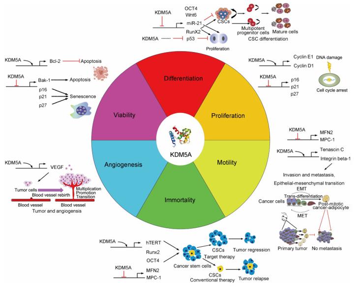 Fig. 1 The biological function and action mechanism of KDM5A.1
Key structural properties of KDM5A:
- The JmjC catalytic domain is responsible for the demethylation function
- The PHD domain recognizes the histone methylation status
- The ARID domain binds to DNA
- The JmjN domain maintains the stability of the enzyme activity
Functions of KDM5A
The main function of KDM5A is to remove the methylation modification of lysine 4 on histone H3, thereby regulating gene transcription. However, it is also involved in various cellular processes, including cell proliferation, differentiation, and DNA damage repair.
| Function | Description |
| Histone demethylation | Specifically removes the H3K4me2/3 methyl marks, inhibiting the expression of target genes. |
| Transcriptional Regulation | By interacting with transcription factors and co-repressor complexes, it regulates the gene expression network. |
| Cell Cycle Regulation | Involved in the transition from G1 to S phase, influencing the cell proliferation process. |
| DNA Damage Response | After DNA damage, it is recruited to the damaged site and participates in the regulation of the repair process. |
| Tumor occurrence and development | It is highly expressed in various cancers, promoting the proliferation, drug resistance and metastasis of cancer cells. |
KDM5A catalyzes demethylation reactions through its JmjC domain. This process relies on iron ions and α-ketoglutarate as cofactors. Its substrate selectivity for different methylation states enables it to have precise targeting in epigenetic regulation.
Applications of KDM5A and KDM5A Antibody in Literature
1. Yang, Guan-Jun, et al. "The emerging role of KDM5A in human cancer." Journal of hematology & oncology 14.1 (2021): 30. https://doi.org/10.1186/s13045-021-01041-1
The research has found that histone methylation is a key modification of chromatin, and its abnormality affects genomic stability and transcriptional regulation. KDM5A, as a histone demethylase, drives the occurrence of various cancers by regulating H3K4 methylation. This article reviews the role of KDM5A in tumors, the progress of inhibitor development, and the future challenges.
2. Troester, Selina, et al. "Transcriptional and epigenetic rewiring by the NUP98:: KDM5A fusion oncoprotein directly activates CDK12." Nature Communications 16.1 (2025): 4656. https://doi.org/10.1038/s41467-025-59930-9
The study found that the NUP98 fusion cancer protein drives childhood acute myeloid leukemia. The study also discovered that NUP98::KDM5A directly regulates key target genes, and through CRISPR screening, CDK12 was identified as the therapeutic target. Inhibiting CDK12 can increase DNA damage and induce the death of leukemia cells.
3. Mitsui, Eishin, et al. "Identification of ryuvidine as a KDM5A inhibitor." Scientific Reports 9.1 (2019): 9952. https://doi.org/10.1038/s41598-019-46346-x
The study found that by using AlphaScreen to screen for KDM5A inhibitors, the active compound ryuvidine was obtained. It can inhibit KDM5A/B/C, increase the level of H3K4me3, and effectively prevent the development of drug resistance in lung cancer PC9 cells, providing a lead compound for targeted KDM5 therapy.
4. Gaillard, Solenne, et al. "KDM5A and KDM5B histone-demethylases contribute to HU-induced replication stress response and tolerance." Biology Open 10.5 (2021): bio057729. https://doi.org/10.1242/bio.057729
The study found that KDM5A/B participates in the replication stress response and tolerance by upregulating RRM2 and maintaining Chk1 activation. KDM5A is enriched at the replication fork and binds to PCNA/Chk1. The study suggests that drugs targeting the enzyme activity may not be able to completely eliminate its oncogenic effect.
5. Peng, Daohu, et al. "Histone demethylase KDM5A promotes tumorigenesis of osteosarcoma tumor." Cell Death Discovery 7.1 (2021): 9. https://doi.org/10.1038/s41420-020-00396-7
The study found that KDM5A is highly expressed in osteosarcoma and is associated with tumor progression. Knockdown of KDM5A can inhibit cell proliferation, induce apoptosis, and regulate P27/Cyclin D1. In vivo experiments confirmed that knocking out KDM5A can inhibit tumor growth, providing a new target for the treatment of osteosarcoma.
Creative Biolabs: KDM5A Antibodies for Research
Creative Biolabs specializes in the production of high-quality KDM5A antibodies for research and industrial applications. Our portfolio includes monoclonal and polyclonal antibodies tailored for ELISA, Flow Cytometry, Western blot, immunohistochemistry, and other diagnostic methodologies.
- Custom KDM5A Antibody Development: Tailor-made solutions to meet specific research requirements.
- Bulk Production: Large-scale antibody manufacturing for industry partners.
- Technical Support: Expert consultation for protocol optimization and troubleshooting.
- Aliquoting Services: Conveniently sized aliquots for long-term storage and consistent experimental outcomes.
For more details on our KDM5A antibodies, custom preparations, or technical support, contact us at email.
Reference
- Yang, Guan-Jun, et al. "The emerging role of KDM5A in human cancer." Journal of hematology & oncology 14.1 (2021): 30. Distributed under Open Access license CC BY 4.0, without modification. https://doi.org/10.1186/s13045-021-01041-1
Anti-KDM5A antibodies
 Loading...


Hot products 
-
Mouse Anti-AGO2 Recombinant Antibody (V2-634169) (CBMAB-AP203LY)

-
Mouse Anti-CD46 Recombinant Antibody (CBFYC-0076) (CBMAB-C0085-FY)

-
Mouse Anti-BIRC7 Recombinant Antibody (88C570) (CBMAB-L0261-YJ)

-
Mouse Anti-CRYAB Recombinant Antibody (A4345) (CBMAB-A4345-YC)

-
Mouse Anti-GFP Recombinant Antibody (28) (CBMAB-G3038-LY)

-
Mouse Anti-ADGRL2 Recombinant Antibody (V2-58519) (CBMAB-L0166-YJ)

-
Mouse Anti-CCS Recombinant Antibody (CBFYC-1093) (CBMAB-C1150-FY)

-
Mouse Anti-COL1A2 Recombinant Antibody (CF108) (V2LY-1206-LY626)

-
Mouse Anti-BLK Recombinant Antibody (CBYY-0618) (CBMAB-0621-YY)

-
Mouse Anti-ELAVL4 Recombinant Antibody (6B9) (CBMAB-1132-YC)

-
Mouse Anti-AP4E1 Recombinant Antibody (32) (CBMAB-A2996-YC)

-
Rat Anti-CD300A Recombinant Antibody (172224) (CBMAB-C0423-LY)

-
Mouse Anti-ENO1 Recombinant Antibody (8G8) (CBMAB-E1329-FY)

-
Mouse Anti-ADGRE5 Recombinant Antibody (V2-360335) (CBMAB-C2088-CQ)

-
Mouse Anti-ABCA3 Recombinant Antibody (V2-178911) (CBMAB-A0145-YC)

-
Rat Anti-ADAM10 Recombinant Antibody (V2-179741) (CBMAB-A1103-YC)

-
Mouse Anti-CASP8 Recombinant Antibody (CBYY-C0987) (CBMAB-C2424-YY)

-
Mouse Anti-ARHGDIA Recombinant Antibody (CBCNA-009) (CBMAB-R0415-CN)

-
Mouse Anti-ATP1B1 Recombinant Antibody (E4) (CBMAB-0463-LY)

-
Mouse Anti-AMH Recombinant Antibody (5/6) (CBMAB-A2527-YC)

- AActivation
- AGAgonist
- APApoptosis
- BBlocking
- BABioassay
- BIBioimaging
- CImmunohistochemistry-Frozen Sections
- CIChromatin Immunoprecipitation
- CTCytotoxicity
- CSCostimulation
- DDepletion
- DBDot Blot
- EELISA
- ECELISA(Cap)
- EDELISA(Det)
- ESELISpot
- EMElectron Microscopy
- FFlow Cytometry
- FNFunction Assay
- GSGel Supershift
- IInhibition
- IAEnzyme Immunoassay
- ICImmunocytochemistry
- IDImmunodiffusion
- IEImmunoelectrophoresis
- IFImmunofluorescence
- IGImmunochromatography
- IHImmunohistochemistry
- IMImmunomicroscopy
- IOImmunoassay
- IPImmunoprecipitation
- ISIntracellular Staining for Flow Cytometry
- LALuminex Assay
- LFLateral Flow Immunoassay
- MMicroarray
- MCMass Cytometry/CyTOF
- MDMeDIP
- MSElectrophoretic Mobility Shift Assay
- NNeutralization
- PImmunohistologyp-Paraffin Sections
- PAPeptide Array
- PEPeptide ELISA
- PLProximity Ligation Assay
- RRadioimmunoassay
- SStimulation
- SESandwich ELISA
- SHIn situ hybridization
- TCTissue Culture
- WBWestern Blot
