PATJ Antibodies
Background
The PATJ gene encodes a scaffold protein present at the tight junctions of cells and is mainly expressed in epithelial cells and neural tissues. This protein interacts with other junction proteins through multiple PDZ domains, jointly maintaining the polarity of cells and the stability of intercellular junction structures, which is crucial for tissue barrier function and cell signal transduction. In 2002, researchers first identified PATJ as an interacting protein of PALS1 through a yeast two-hybrid system. Subsequent studies gradually revealed its core role in the apical junction complex of cells. Mutations in this gene are associated with hereditary sensorineural deafness and skin barrier dysfunction. Its highly modular domain design has become a classic model for studying cell junction assembly and protein interaction networks, providing an important basis for revealing the mechanism of epithelial polarity regulation and the pathology of related diseases.
Structure of PATJ
PATJ is a large scaffold protein, and its molecular weight varies with different splicing variants. The molecular weight of the main human isomer is approximately 220 kDa. This protein contains multiple highly conserved domains, and its composition varies significantly among different species.
| Species | Human | Mouse | Zebrafish |
| Molecular Weight (kDa) | ~220 | ~215 | ~190 |
| Primary Structural Differences | Contains 10 PDZ domains and 1 L27 domain | The number of PDZ domains is highly consistent with that of humans | The number of PDZ domains is relatively small and they are core functional areas |
The primary structure of the PATJ protein is composed of multiple PDZ domains linearly connected in series, forming a modular architecture. Its secondary structure is mainly composed of β -folds and α -helices within each PDZ domain, which jointly form the protein binding interface. The key L27 domain binds to chaperone proteins such as PALS1 through helical interactions at the amino terminus, while the continuous PDZ domain assemps into a macroscopic polar protein network at the cell apex by binding to the carboxyl terminus of transmembrane proteins (such as CRB3 and Claudins).
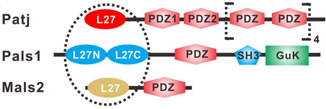 Fig. 1 Schematic of the domain organization of rat Patj.1
Key structural properties of PATJ:
- Series connection of modular PDZ domains
- N-terminal domain L27 structure mediated starting complex assembly
- Lacks typical enzymatic catalytic activity
Functions of PATJ
The core function of the PATJ protein is to maintain cell polarity and the assembly of junction complexes, while also participating in the regulation of various cellular physiological processes.
| Function | Description |
| Maintain cell polarity | By binding multiple PDZ domains with transmembrane proteins and signaling molecules, the apic-basal polarity of epithelial cells is established and stabilized. |
| Connection complex assembly | As a scaffold protein, it collaborates with proteins such as PALS1 and CRB3 to form tight junction complexes at the cell top, ensuring intercellular adhesion and barrier functions. |
| Cell signaling | By recruiting signal molecules to specific membrane regions, it participates in the regulation of signaling pathways related to cell proliferation and differentiation, such as Hippo and YAP. |
| Establishment of organizational barriers | In epithelial and endothelial tissues, it directly participates in the formation of physical barriers and selective permeability barriers by stabilizing tight junction structures. |
| Association of disease occurrence | The dysfunction is closely related to sensorineural deafness, skin barrier defect, polarity loss and invasive enhancement of some cancers. |
PATJ simultaneously binds multiple ligands in a low-affinity and high-specificity manner through its highly modular PDZ domain array. This multivalent interaction mode enables it to efficiently assemble large protein networks. Compared with a single high-affinity interaction, it is more suitable for performing complex functions that require spatial coordination, such as establishing cell polarity.
Applications of PATJ and PATJ Antibody in Literature
1. Zhang J, Yang X,et al. "Structure of an L27 domain heterotrimer from cell polarity complex Patj/Pals1/Mals2 reveals mutually independent L27 domain assembly mode." Journal of Biological Chemistry 287.14 (2012): 11132-11140. https://doi.org/10.1074/jbc.M111.321216
This article reveals the molecular mechanism by which the scaffold protein PATJ forms a heterotrimer with PALS1/MALS2 through tandem L27 domain. Structural analysis and biochemical experiments indicated that the two pairs of L27 heterodimers in this complex assembled independently of each other. This novel assembly pattern provides new insights into the regulation of cell polarity.
2. Medina-Dols, Aina, et al. "Role of PATJ in stroke prognosis by modulating endothelial to mesenchymal transition through the Hippo/Notch/PI3K axis." Cell death discovery 10.1 (2024): 85. https://doi.org/10.1038/s41420-024-01857-z
This article reveals that genome-wide association studies have found that PATJ is associated with functional recovery after ischemic stroke. Patient data show that the down-regulation of PATJ expression 24 hours after stroke is associated with a good prognosis. Mechanologically, mouse and cell experiments have shown that ischemia and hypoxia lead to a reduction in PATJ, which in turn induces endothelial-interstitial transition in endothelial cells through signaling pathways such as MUC1-C. This may be an early beneficial step to promote post-stroke recovery.
3. Fiedler, Julia, et al. "PATJ inhibits histone deacetylase 7 to control tight junction formation and cell polarity." Cellular and Molecular Life Sciences 80.11 (2023): 333. https://doi.org/10.1007/s00018-023-04994-3
This study reveals a new mechanism by which PATJ maintains the polarity of epithelial cells by regulating HDAC7. The deletion of PATJ can disrupt cell polarity and lumen formation. The mechanism lies in the fact that PATJ can bind to and inhibit HDAC7, thereby regulating the expression of genes related to cell connection. This function is independent of its classic partner Pals1, revealing a new role of PATJ at the transcriptional regulatory level.
4. Epting, Daniel, et al. "PATJ deficiency leads to cystic kidney disease and related ciliopathies." Human Genetics and Genomics Advances 7.1 (2026). https://doi.org/10.1016/j.xhgg.2025.100514
In this study, through gene sequencing, a biallelic truncation mutation of the PATJ gene was first identified as the cause of the disease in a ciliatosis fetus with renal cysts and hydrocephalus. The zebrafish model was used to verify that the absence of PATJ would lead to a typical ciliary phenotype, revealing the previously unknown key role of PATJ in ciliary formation and function, and adding a new member to the ciliary disease family.
5. Gu, Jiayu, et al. "Isolation and Characterization of a Lytic Phage PaTJ Against Pseudomonas aeruginosa." Viruses 16.12 (2024): 1816. https://doi.org/10.3390/v16121816
In this study, a novel Pseudomonas lysis phage, PaTJ, was isolated from wastewater. This bacteriophage uses type IV fimbriae as receptors and is characterized by a short incubation period and a large outbreak volume. Transcriptome analysis revealed the gene expression pattern in the early stage of infection and confirmed that PaTJ can effectively degrade biofilms and has application potential in the treatment of drug-resistant bacterial infections.
Creative Biolabs: PATJ Antibodies for Research
Creative Biolabs specializes in the production of high-quality PATJ antibodies for research and industrial applications. Our portfolio includes monoclonal antibodies tailored for ELISA, Flow Cytometry, Western blot, immunohistochemistry, and other diagnostic methodologies.
- Custom PATJ Antibody Development: Tailor-made solutions to meet specific research requirements.
- Bulk Production: Large-scale antibody manufacturing for industry partners.
- Technical Support: Expert consultation for protocol optimization and troubleshooting.
- Aliquoting Services: Conveniently sized aliquots for long-term storage and consistent experimental outcomes.
For more details on our PATJ antibodies, custom preparations, or technical support, contact us at email.
Reference
- Zhang J, Yang X,et al. "Structure of an L27 domain heterotrimer from cell polarity complex Patj/Pals1/Mals2 reveals mutually independent L27 domain assembly mode." Journal of Biological Chemistry 287.14 (2012): 11132-11140. https://doi.org/10.1074/jbc.M111.321216
Anti-PATJ antibodies
 Loading...


Hot products 
-
Mouse Anti-AKR1C3 Recombinant Antibody (V2-12560) (CBMAB-1050-CN)

-
Mouse Anti-AGO2 Recombinant Antibody (V2-634169) (CBMAB-AP203LY)

-
Mouse Anti-CD19 Recombinant Antibody (CBXC-1224) (CBMAB-C1491-CQ)

-
Mouse Anti-ABCA3 Recombinant Antibody (V2-178911) (CBMAB-A0145-YC)

-
Mouse Anti-APP Recombinant Antibody (DE2B4) (CBMAB-1122-CN)

-
Mouse Anti-ADAM12 Recombinant Antibody (V2-179752) (CBMAB-A1114-YC)

-
Mouse Anti-BACE1 Recombinant Antibody (61-3E7) (CBMAB-1183-CN)

-
Mouse Anti-FPR2 Recombinant Antibody (1D6) (CBMAB-F2628-CQ)

-
Mouse Anti-dsRNA Recombinant Antibody (2) (CBMAB-D1807-YC)

-
Mouse Anti-CARD11 Recombinant Antibody (CBFYC-0811) (CBMAB-C0866-FY)

-
Mouse Anti-BIRC3 Recombinant Antibody (16E63) (CBMAB-C3367-LY)

-
Mouse Anti-CD24 Recombinant Antibody (2Q1282) (CBMAB-C1624-CN)

-
Mouse Anti-DLG1 Monolconal Antibody (4F3) (CBMAB-0225-CN)

-
Mouse Anti-CASP7 Recombinant Antibody (10-01-62) (CBMAB-C2005-LY)

-
Mouse Anti-DISP2 Monoclonal Antibody (F66A4B1) (CBMAB-1112CQ)

-
Mouse Anti-CORO1A Recombinant Antibody (4G10) (V2LY-1206-LY806)

-
Mouse Anti-CD33 Recombinant Antibody (6C5/2) (CBMAB-C8126-LY)

-
Mouse Anti-CD83 Recombinant Antibody (HB15) (CBMAB-C1765-CQ)

-
Rabbit Anti-DLK1 Recombinant Antibody (9D8) (CBMAB-D1061-YC)

-
Mouse Anti-BBS2 Recombinant Antibody (CBYY-0253) (CBMAB-0254-YY)

- AActivation
- AGAgonist
- APApoptosis
- BBlocking
- BABioassay
- BIBioimaging
- CImmunohistochemistry-Frozen Sections
- CIChromatin Immunoprecipitation
- CTCytotoxicity
- CSCostimulation
- DDepletion
- DBDot Blot
- EELISA
- ECELISA(Cap)
- EDELISA(Det)
- ESELISpot
- EMElectron Microscopy
- FFlow Cytometry
- FNFunction Assay
- GSGel Supershift
- IInhibition
- IAEnzyme Immunoassay
- ICImmunocytochemistry
- IDImmunodiffusion
- IEImmunoelectrophoresis
- IFImmunofluorescence
- IGImmunochromatography
- IHImmunohistochemistry
- IMImmunomicroscopy
- IOImmunoassay
- IPImmunoprecipitation
- ISIntracellular Staining for Flow Cytometry
- LALuminex Assay
- LFLateral Flow Immunoassay
- MMicroarray
- MCMass Cytometry/CyTOF
- MDMeDIP
- MSElectrophoretic Mobility Shift Assay
- NNeutralization
- PImmunohistologyp-Paraffin Sections
- PAPeptide Array
- PEPeptide ELISA
- PLProximity Ligation Assay
- RRadioimmunoassay
- SStimulation
- SESandwich ELISA
- SHIn situ hybridization
- TCTissue Culture
- WBWestern Blot
