RHOB Antibodies
Background
RHOB gene encodes a small GTP binding protein, which is a member of the Rho family and is widely present in eukaryotic cells. This protein participates in important physiological processes such as cell migration, proliferation and apoptosis by regulating cytoskeletal recombination and cell signal transduction pathways. Studies have shown that RHOB plays a dual role in tumorigenesis and development. It can not only act as a tumor suppressor gene to induce apoptosis but also promote tumor metastasis, making it a key target in cancer research. This gene was first identified by Alan Hall's team in 1985. Its unique membrane localization characteristics and GTPase activity provide key clues for understanding the mechanism of cell movement. In recent years, scientists have deeply analyzed the three-dimensional structure of RHOB and its interaction mechanism with effectors through techniques such as cryo-electron microscopy. These discoveries have greatly promoted the development of cell biology and tumor treatment fields.
Structure of RHOB
RHOB is a small-molecular-weight GTP-binding protein with a molecular weight of approximately 21.5 kDa and is highly conserved across different species. Its molecular weight may vary slightly due to post-translational modifications such as isoprene modification.
| Species | Human | Mouse | Rat | Fruit fly |
| Molecular Weight (kDa) | 21.5 | 21.4 | 21.5 | 20.8 |
| Primary Structural Differences | C-terminal CAAX motif (isoprene site) | Highly conserved, with over 90% homology of RHOB in humans | Similar to humans, it regulates the cytoskeleton | Participate in developmental regulation |
RHOB is composed of 193 amino acids and has a typical GTPase domain, regulating the GTP/GDP binding state through the Switch I and Switch II regions. The C-terminal CAAX motif has been modified by isoprene, which promotes membrane localization and participates in signal transduction. The tertiary structure of RHOB consists of 6 β -folded sheets and 5 α -helices, forming GTP binding pockets. Key residues such as Gly14, Thr19 and Gln63 are involved in the hydrolysis of GTP, while Thr37 and Asp65 regulate the interaction of effector proteins. This protein plays a significant role in cancer and immune responses by regulating actin kinetics and cell migration.
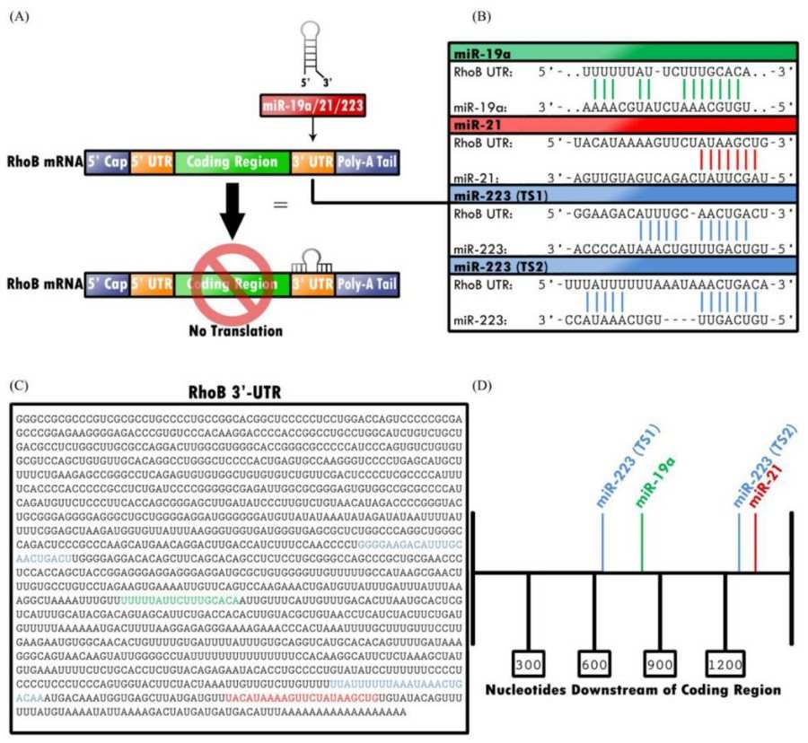 Fig. 1 Translation of RHOB is epigenetically downregulated by miRNA-19, -21, and -223.1
Key structural properties of RHOB:
- GTPase domain
- Switch I/II variable zone
- C-end CAAX motif
- Effector protein binding region
- Key amino acid residues
Functions of RHOB
The core function of the RHOB gene is to regulate cytoskeletal recombination and signal transduction, and it also plays a key role in various pathophysiological processes.
| Function | Description |
| Cytoskeleton regulation | By activating effector proteins such as ROCK and mDia, it regulates actin polymerization and stress fiber formation. |
| Cell migration control | Coordinating the dynamic changes of the frontier membrane folds and adhesion spots of cells affects tumor metastasis and wound healing. |
| Cell cycle regulation | Through target genes such as p21 and cyclin D1, it affects G1/S phase transition. |
| Dual effects of tumors | Inhibits proliferation in some cancers (e.g., lung cancer) and promotes invasion in others (e.g., glioma). |
| Angiogenesis regulation | Endothelial cell migration and blood vessel formation are affected by VEGF signaling pathway. |
| Stress response | Respond to DNA damage and oxidative stress, and participate in apoptosis or survival decisions. |
The activity of RHOB is strictly regulated by the GTP/GDP conversion, and its effect shows significant cell type and microenvironment dependence. Unlike other family members such as RhoA, RHOB is often in a continuously activated state in resting cells. This unique regulatory pattern makes it a key "molecular switch" for cellular homeostasis.
Applications of RHOB and RHOB Antibody in Literature
1. Yang, Jianming, et al. "RhoB affects colitis through modulating cell signaling and intestinal microbiome." Microbiome 10.1 (2022): 149. https://doi.org/10.1186/s40168-022-01347-3
Research has found that RhoB is significantly elevated in patients with ulcerative colitis (UC) and mice with DSS-induced colitis. Reducing RhoB expression can alleviate colitis symptoms, promote goblet cell differentiation and epithelial regeneration, inhibit the Wnt pathway and activate the p38 MAPK pathway, while increasing short-chain fatty acid-producing bacteria and their concentrations. RhoB may serve as a biomarker and therapeutic target for UC.
2. Ju, Julia A., and Daniele M. Gilkes. "RhoB: team oncogene or team tumor suppressor?." Genes 9.2 (2018): 67. https://doi.org/10.3390/genes9020067
Research has found that RhoB, unlike RhoA/RhoC, has a dual role in tumors, capable of both promoting and inhibiting cancer. Its unique C-terminal modification and subcellular localization (such as in endosomes and nuclei) make its function complex, and it may play the opposite role of RhoA/RhoC in the tumor microenvironment.
3. Gu, Jianyou, et al. "Hsa-miR-3178/RhoB/PI3K/Akt, a novel signaling pathway regulates ABC transporters to reverse gemcitabine resistance in pancreatic cancer." Molecular Cancer 21.1 (2022): 112. https://doi.org/10.1186/s12943-022-01587-9
Studies have found that hsa-miR-3178 promotes resistance to gemcitabine in pancreatic cancer by targeting and inhibiting RhoB, activating the PI3K/Akt pathway and upregulating the ABC transporter (P-gp/BCRP/MRP1). Low expression of RhoB indicates a poor prognosis for patients. Restoring RhoB can reverse drug resistance, suggesting that the miR-3178-RhoB axis may become a new target for overcoming chemotherapy resistance.
4. Bailey, Cedric AR. "Regulation of RhoB gene expression during tumorigenesis and aging process and Its potential applications in these processes."Cancers 11.6 (2019): 818. https://doi.org/10.3390/cancers11060818
Research has found that RhoB, as a member of the Rho GTPase family, affects cell function by regulating pathways such as EGFR/Ras/PI3K-Akt/mTOR. Its expression is regulated by HDAC and miRNAs and plays a tissue-specific role in tumorigenesis. The down-regulation of RhoB is associated with tumor development, and restoring its expression may become a new strategy for cancer prevention and treatment.
5. Kopsida, Maria, et al. "RhoB expression associated with chemotherapy response and prognosis in colorectal cancer." Cancer Cell International 24.1 (2024): 75. https://doi.org/10.1186/s12935-024-03236-1
Studies have found that high expression of RhoB reduces the chemosensitivity of colorectal cancer to 5-FU and oxaliplatin and is associated with a poor prognosis for patients. Gene knockout of RhoB can inhibit tumor migration, promote apoptosis and regulate autophagy. The mechanism involves caspase-3 activation and ROS regulation. Targeted inhibition of RhoB may enhance the effect of chemotherapy.
Creative Biolabs: RHOB Antibodies for Research
Creative Biolabs specializes in the production of high-quality RHOB antibodies for research and industrial applications. Our portfolio includes monoclonal antibodies tailored for ELISA, Flow Cytometry, Western blot, immunohistochemistry, and other diagnostic methodologies.
- Custom RHOB Antibody Development: Tailor-made solutions to meet specific research requirements.
- Bulk Production: Large-scale antibody manufacturing for industry partners.
- Technical Support: Expert consultation for protocol optimization and troubleshooting.
- Aliquoting Services: Conveniently sized aliquots for long-term storage and consistent experimental outcomes.
For more details on our RHOB antibodies, custom preparations, or technical support, contact us at email.
Reference
- Bailey, Cedric AR. "Regulation of RhoB gene expression during tumorigenesis and aging process and Its potential applications in these processes."Cancers 11.6 (2019): 818. https://doi.org/10.3390/cancers11060818
Anti-RHOB antibodies
 Loading...


Hot products 
-
Mouse Anti-CD63 Recombinant Antibody (CBXC-1200) (CBMAB-C1467-CQ)

-
Mouse Anti-8-oxoguanine Recombinant Antibody (V2-7697) (CBMAB-1869CQ)

-
Mouse Anti-CD247 Recombinant Antibody (6B10.2) (CBMAB-C1583-YY)

-
Mouse Anti-ATP1A2 Recombinant Antibody (M7-PB-E9) (CBMAB-A4013-YC)

-
Mouse Anti-AMACR Recombinant Antibody (CB34A) (CBMAB-CA034LY)

-
Mouse Anti-BACE1 Recombinant Antibody (CBLNB-121) (CBMAB-1180-CN)

-
Mouse Anti-ELAVL4 Recombinant Antibody (6B9) (CBMAB-1132-YC)

-
Mouse Anti-GFAP Recombinant Antibody (24) (CBMAB-G2927-LY)

-
Rat Anti-ABCC11 Recombinant Antibody (V2-179001) (CBMAB-A0236-YC)

-
Mouse Anti-CD24 Recombinant Antibody (HIS50) (CBMAB-C10123-LY)

-
Mouse Anti-CECR2 Recombinant Antibody (CBWJC-2465) (CBMAB-C3533WJ)

-
Mouse Anti-AQP2 Recombinant Antibody (E-2) (CBMAB-A3358-YC)

-
Mouse Anti-FOXA3 Recombinant Antibody (2A9) (CBMAB-0377-YC)

-
Mouse Anti-CORO1A Recombinant Antibody (4G10) (V2LY-1206-LY806)

-
Mouse Anti-ENO1 Recombinant Antibody (8G8) (CBMAB-E1329-FY)

-
Mouse Anti-ACTG1 Recombinant Antibody (V2-179597) (CBMAB-A0916-YC)

-
Rat Anti-EMCN Recombinant Antibody (28) (CBMAB-E0280-FY)

-
Mouse Anti-ADRB2 Recombinant Antibody (V2-180026) (CBMAB-A1420-YC)

-
Rabbit Anti-ABL1 (Phosphorylated Y245) Recombinant Antibody (V2-505716) (PTM-CBMAB-0465LY)

-
Mouse Anti-dsRNA Recombinant Antibody (2) (CBMAB-D1807-YC)

- AActivation
- AGAgonist
- APApoptosis
- BBlocking
- BABioassay
- BIBioimaging
- CImmunohistochemistry-Frozen Sections
- CIChromatin Immunoprecipitation
- CTCytotoxicity
- CSCostimulation
- DDepletion
- DBDot Blot
- EELISA
- ECELISA(Cap)
- EDELISA(Det)
- ESELISpot
- EMElectron Microscopy
- FFlow Cytometry
- FNFunction Assay
- GSGel Supershift
- IInhibition
- IAEnzyme Immunoassay
- ICImmunocytochemistry
- IDImmunodiffusion
- IEImmunoelectrophoresis
- IFImmunofluorescence
- IGImmunochromatography
- IHImmunohistochemistry
- IMImmunomicroscopy
- IOImmunoassay
- IPImmunoprecipitation
- ISIntracellular Staining for Flow Cytometry
- LALuminex Assay
- LFLateral Flow Immunoassay
- MMicroarray
- MCMass Cytometry/CyTOF
- MDMeDIP
- MSElectrophoretic Mobility Shift Assay
- NNeutralization
- PImmunohistologyp-Paraffin Sections
- PAPeptide Array
- PEPeptide ELISA
- PLProximity Ligation Assay
- RRadioimmunoassay
- SStimulation
- SESandwich ELISA
- SHIn situ hybridization
- TCTissue Culture
- WBWestern Blot
