CD63 Antibodies
Background
CD63 is a tetraspanin protein that is widely distributed in endosomes and lysosomes and is a member of the Tetraspanin protein family. This protein regulates intracellular material transport and intercellular communication by participating in membrane transport and vesicle formation processes, especially playing a key role in multivesicular body (MVBs) generation and exosome release. First identified in platelet activation studies in the 1980s, CD63 was later discovered to be one of the classic markers of exosomes, and its expression level is often used as an important basis for exosome identification. The topological structure and functional domain interaction mechanism of this protein have been widely studied, providing an important molecular basis for understanding intracellular membrane transport, cellular signal transduction, and the regulation of the tumor immune microenvironment.
Structure of CD63
CD63 is a transmembrane protein with a molecular weight of approximately 25-50 kDa, and its specific molecular weight varies depending on the degree of glycosylation modification. This protein belongs to the tetraspanin superfamily and has a highly conserved structural core in different species.
| Species | Human | Mouse | Rat | Bovine |
| Molecular Weight (kDa) | 25-50 | 25-50 | 25-50 | 25-50 |
| Primary Structural Differences | Highly glycosylated | The N-glycosylation sites are different | Conservative intracellular area | The C-terminal sequence has slight variations |
The CD63 protein is composed of 239 amino acids, forming four highly conserved transmembrane domains (TM1-TM4), two extracellular loops (EC1, EC2), and the intracellular N-terminus and C-terminus. Among them, the larger second extracellular loop (EC2) contains a characteristic "CCG" motif and conserved disulfide bonds, which are involved in protein-protein interactions. This protein regulates membrane protein transport and cellular signal transduction by forming the tetraspanin web. Its intracellular distribution is mainly in the endosome/lysosomal system and is widely used as a marker of exosomes.
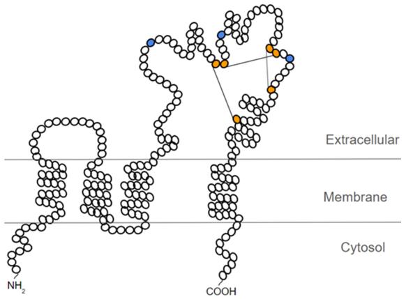 Fig. 1 Schematic representation of the probable structure of CD63.1
Key structural properties of CD63:
- Four transmembrane domain structure formed four typical polymers conformation
- Extracellular the second (EC2) including conservative CCG die bodies and disulfide bond
- Intracellular C tail including lysosomal targeting signal
Functions of CD63
The main function of the CD63 protein is to participate in intracellular transport and exosome formation. However, it also involves a variety of cellular processes, including cell signaling and membrane protein organization.
| Function | Description |
| Intracellular transport | CD63 regulates the material transport process from endosomes to lysosomes, affecting the sorting and degradation of proteins. |
| Exosome biogenesis | As a landmark protein of exosomes, it participates in the formation of multivesicular bodies (MVB) and the release of exosomes. |
| Cell adhesion and migration | Through the tetraspanin network with other membrane protein interactions, affect the cell adhesion, movement, and signal transduction. |
| Immune regulation | In antigen-presenting cells participate in MHC class II molecules transport and the regulation of immune response. |
| Virus infection and host defense | Some viruses use CD63 to enter or exit host cells, and at the same time, it is also involved in the antiviral response of cells. |
CD63 interacts specifically with various ligands and membrane proteins through its extracellular domain to form functional complexes, thereby widely participating in cell membrane compartmentalization and transmembrane signal transduction events.
Applications of CD63 and CD63 Antibody in Literature
1. Wang, Xinyue, et al. "A novel rabbit anti-myoglobin monoclonal antibody's potential application in rhabdomyolysis associated acute kidney injury." International Journal of Molecular Sciences 24.9 (2023): 7822. https://doi.org/10.3390/v13040675
The article indicates that CD63 is a quadruple transmembrane protein that participates in endocytic transport and extracellular vesicle cargo sorting. Research has found that LMP1 can alter the interaction group of CD63, shifting it from transport function to metabolic and translation processes, and may form complexes with mTOR and others to regulate signal transduction.
2. Odaka, Haruki, et al. "CD63-positive extracellular vesicles are potential diagnostic biomarkers of pancreatic ductal adenocarcinoma." BMC gastroenterology 22.1 (2022): 153. https://doi.org/10.1186/s12876-022-02228-7
The article indicates that the level of CD63+ extracellular vesicles in the serum of patients with pancreatic ductal adenocarcinoma (PDAC) is significantly elevated, demonstrating excellent diagnostic performance (AUC: 0.846), especially when combined with CA19-9 in the early stage (AUC: 0.903), and the level decreases after surgery, showing its potential as a biomarker for PDAC.
3. Danquah, Bright D., et al. "Mass Spectrometric analysis of antibody—Epitope peptide complex dissociation: Theoretical concept and practical procedure of binding strength characterization." Molecules 25.20 (2020): 4776. https://doi.org/10.1038/s41598-021-99777-w
The article indicates that the level of CD63 in plasma exosomes is significantly elevated in patients with sepsis, positively correlated with the SOFA score. Moreover, a high level of CD63 (>126 µg/mL) can predict 28-day in-hospital mortality and 90-day survival, suggesting its potential as a biomarker for evaluating the severity and prognosis of sepsis.
4. Justo, Beatriz Laís, and Miriam Galvonas Jasiulionis. "Characteristics of TIMP1, CD63, and β1-integrin and the functional impact of their interaction in cancer." International journal of molecular sciences 22.17 (2021): 9319. https://doi.org/10.3390/ijms22179319
The article indicates that TIMP-1 can form a membrane complex with CD63 and β 1-integrin. This complex not only inhibits the activity of metalloproteinases but also participates in regulating cell growth and survival signaling pathways. Moreover, it is highly expressed in various tumors and may be associated with a poor prognosis. Its assembly and function are regulated by N-glycosylation.
5. Fan, Yé, et al. "Differential proteomics argues against a general role for CD9, CD81 or CD63 in the sorting of proteins into extracellular vesicles." Journal of Extracellular Vesicles 12.8 (2023): 12352. https://doi.org/10.1002/jev2.12352
The article indicates that CD63, CD9 and CD81 are the main marker proteins of extracellular vesicles (EVs), but in MCF7 cells, their influence on the protein composition of EVs is limited. The co-deletion of CD9 and CD81 will significantly reduce the contents of their ligands CD9P-1 and EWI-2 in EVs. These four-transmembrane proteins are more involved in regulating the expression and transport of related proteins rather than directly determining the composition of EVs.
Creative Biolabs: CD63 Antibodies for Research
Creative Biolabs specializes in the production of high-quality CD63 antibodies for research and industrial applications. Our portfolio includes monoclonal antibodies tailored for ELISA, Flow Cytometry, Western blot, immunohistochemistry, and other diagnostic methodologies.
- Custom CD63 Antibody Development: Tailor-made solutions to meet specific research requirements.
- Bulk Production: Large-scale antibody manufacturing for industry partners.
- Technical Support: Expert consultation for protocol optimization and troubleshooting.
- Aliquoting Services: Conveniently sized aliquots for long-term storage and consistent experimental outcomes.
For more details on our CD63 antibodies, custom preparations, or technical support, contact us at email.
Reference
- Justo, Beatriz Laís, and Miriam Galvonas Jasiulionis. "Characteristics of TIMP1, CD63, and β1-integrin and the functional impact of their interaction in cancer." International journal of molecular sciences 22.17 (2021): 9319. https://doi.org/10.3390/ijms22179319
Anti-CD63 antibodies
 Loading...


Hot products 
-
Mouse Anti-DISP2 Monoclonal Antibody (F66A4B1) (CBMAB-1112CQ)

-
Mouse Anti-APOE Recombinant Antibody (A1) (CBMAB-0078CQ)

-
Mouse Anti-CD24 Recombinant Antibody (HIS50) (CBMAB-C10123-LY)

-
Mouse Anti-CRTAM Recombinant Antibody (CBFYC-2235) (CBMAB-C2305-FY)

-
Rabbit Anti-ALDOA Recombinant Antibody (D73H4) (CBMAB-A2314-YC)

-
Mouse Anti-AGK Recombinant Antibody (V2-258056) (CBMAB-M0989-FY)

-
Mouse Anti-ACTB Recombinant Antibody (V2-179553) (CBMAB-A0870-YC)

-
Rat Anti-CD34 Recombinant Antibody (MEC 14.7) (CBMAB-C10196-LY)

-
Mouse Anti-CCL18 Recombinant Antibody (64507) (CBMAB-C7910-LY)

-
Mouse Anti-GDF5 Recombinant Antibody (1F4) (CBMAB-G2740-LY)

-
Mouse Anti-CCT6A/B Recombinant Antibody (CBXC-0168) (CBMAB-C5570-CQ)

-
Rabbit Anti-ATF4 Recombinant Antibody (D4B8) (CBMAB-A3872-YC)

-
Mouse Anti-B2M Recombinant Antibody (CBYY-0050) (CBMAB-0050-YY)

-
Mouse Anti-FLI1 Recombinant Antibody (CBXF-0733) (CBMAB-F0435-CQ)

-
Mouse Anti-CTCF Recombinant Antibody (CBFYC-2371) (CBMAB-C2443-FY)

-
Rat Anti-(1-5)-α-L-Arabinan Recombinant Antibody (V2-501861) (CBMAB-XB0003-YC)

-
Mouse Anti-GFAP Recombinant Antibody (24) (CBMAB-G2927-LY)

-
Mouse Anti-DDC Recombinant Antibody (8E8) (CBMAB-0992-YC)

-
Mouse Anti-AP4E1 Recombinant Antibody (32) (CBMAB-A2996-YC)

-
Rat Anti-CD300A Recombinant Antibody (172224) (CBMAB-C0423-LY)

- AActivation
- AGAgonist
- APApoptosis
- BBlocking
- BABioassay
- BIBioimaging
- CImmunohistochemistry-Frozen Sections
- CIChromatin Immunoprecipitation
- CTCytotoxicity
- CSCostimulation
- DDepletion
- DBDot Blot
- EELISA
- ECELISA(Cap)
- EDELISA(Det)
- ESELISpot
- EMElectron Microscopy
- FFlow Cytometry
- FNFunction Assay
- GSGel Supershift
- IInhibition
- IAEnzyme Immunoassay
- ICImmunocytochemistry
- IDImmunodiffusion
- IEImmunoelectrophoresis
- IFImmunofluorescence
- IGImmunochromatography
- IHImmunohistochemistry
- IMImmunomicroscopy
- IOImmunoassay
- IPImmunoprecipitation
- ISIntracellular Staining for Flow Cytometry
- LALuminex Assay
- LFLateral Flow Immunoassay
- MMicroarray
- MCMass Cytometry/CyTOF
- MDMeDIP
- MSElectrophoretic Mobility Shift Assay
- NNeutralization
- PImmunohistologyp-Paraffin Sections
- PAPeptide Array
- PEPeptide ELISA
- PLProximity Ligation Assay
- RRadioimmunoassay
- SStimulation
- SESandwich ELISA
- SHIn situ hybridization
- TCTissue Culture
- WBWestern Blot
