COL5A1 Antibodies
Background
The COL5A1 gene encodes the α1 chain of type V collagen, which is an important component of fibrous collagen in the extracellular matrix and is mainly distributed in connective tissues such as skin, tendons, and blood vessels. Its expression products form heterotrimers and are jointly assembled with type I collagen to form a fibrous network, which not only provides mechanical support for tissues but also affects tissue toughness by regulating the diameter and spatial arrangement of fibers. Mutations in this gene can disrupt the normal morphology of collagen fibers, leading to the classic occurrence of Ellers-Danlos syndrome, which is clinically manifested as excessive skin extension, excessive joint movement and tissue fragility. As a key molecule for maintaining tissue integrity, COL5A1 has become an important target for the mechanism research and clinical diagnosis of connective tissue diseases, and its molecular pathological research continuously promotes the development of related precision medical strategies.
Structure of COL5A1
The V-type collagen α1 chain encoded by the COL5A1 gene is a high-molecular-weight protein with a molecular weight of approximately 183 kDa. The molecular weight of this protein may vary slightly among different species due to selective splicing of transcripts and the degree of post-translational modification.
| Species | Human | Mouse | Bovine | Rat | Chicken |
| Molecular Weight (kDa) | 183 | 182.5 | 183.2 | 182.8 | 181.9 |
| Primary Structural Differences | Contains an N-terminal propeptide and a long collagen domain | Collagen structure domain highly conservative | There are differences in glycosylation sites | The sequences of the triple helix regions are similar | Structure of domain is relatively primitive |
The COL5A1 protein is composed of approximately 1,800 amino acid residues. Its typical structure includes an N-terminal signal peptide, a propeptide domain, and a collagen triple helix repeat sequence of up to 1,500 amino acids (Gly-X-Y). This protein mediates inter-chain recognition through its C-terminal non-collagen domain and assembles with the COL5A2 and COL5A3 chains to form a V-type collagen heterotrimer. The glycine residues (Gly) that must appear at every two positions in the triple helix structure form the basis of structural stability, while the regular arrangement of proline (X position) and hydroxyproline (Y position) further enhances the rigidity of the triple helix structure through a hydrogen bond network. This highly ordered secondary structure provides a crucial mechanical support function for the extracellular matrix.
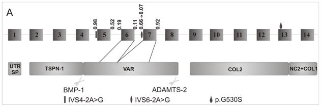 Fig. 1 Exon and domain structure of the COL5A1 N-propeptide encoding region.1
Key structural properties of COL5A1:
- Characteristic Gly-X-Y triple helix repeat domain
- Conserved N-terminal propeptide autoregulatory region
- Form the C-terminal non-collagen domain of the heterotrimer
Functions of COL5A1
The core function of the protein encoded by the COL5A1 gene is to form the V-type collagen fiber network in the extracellular matrix. Its main functions include:
| Function | Description |
| Regulation of fiber formation | Co-assembled with type I collagen, it regulates the diameter and spatial arrangement of fibrils and affects the mechanical properties of tissues. |
| The role of matrix scaffolds | 3D networks are formed in connective tissues such as skin and tendons to provide structural support and attachment sites for cells. |
| Developmental regulatory function | By influencing the signal transduction, and extracellular matrix in tissue morphogenesis in the process of embryonic development. |
| Wound repair support | After the tissue damage to provide temporary support for cell migration, promote orderly repair process. |
| Regulation of cell behavior | Through the interaction between integrin affect cell proliferation, differentiation and apoptosis, etc. |
Unlike type I collagen, which mainly provides mechanical support, type V collagen plays a specific role in regulating fiber morphogenesis and maintaining tissue homeostasis through its unique surface distribution and proteoglycan binding ability. This functional characteristic makes it a key regulatory factor for connective tissue integrity.
Applications of COL5A1 and COL5A1 Antibody in Literature
1. Gu, Sujie, et al. "COL5A1 serves as a biomarker of tumor progression and poor prognosis and may be a potential therapeutic target in gliomas." Frontiers in oncology 11 (2021): 752694. https://doi.org/10.3389/fonc.2021.752694
This study confirmed that COL5A1 is highly expressed in glioma and is associated with malignant progression and poor prognosis. Inhibiting COL5A1 can reduce tumor proliferation and migration and enhance temozolomide sensitivity. COL5A1 also affects the tumor immune microenvironment through dendritic cells and is a potential therapeutic target.
2. Pabalan, Noel, et al. "Association of COL5A1 gene polymorphisms and risk of tendon-ligament injuries among Caucasians: a meta-analysis." Sports Medicine-Open 4.1 (2018): 46. https://doi.org/10.1186/s40798-018-0161-0
This study confirmed that COL5A1 is highly expressed in glioma, indicating a poor prognosis. Inhibiting its expression can reduce the proliferation and migration of cancer cells and enhance chemotherapy sensitivity, making it a potential therapeutic target.
3. Sun, Zhuo, Paweł Cięszczyk, and Aleksandra Bojarczuk. "COL5A1 rs13946 Polymorphism and Anterior Cruciate Ligament Injury: Systematic Review and Meta-Analysis." International Journal of Molecular Sciences 26.13 (2025): 6340. https://doi.org/10.3390/ijms26136340
This study confirmed that the meta-analysis indicated that the rs13946 polymorphism of the COL5A1 gene was associated with the risk of anterior cruciate ligament injury. In the Caucasian population, carrying the C allele or C/- genotype may have a protective effect on ACL injury.
4. Dalewski, Bartosz, et al. "COL5A1 RS12722 is associated with temporomandibular joint anterior disc displacement without reduction in polish caucasians." Cells 10.9 (2021): 2423. https://doi.org/10.3390/cells10092423
Research has found that the rs12722 polymorphism of the COL5A1 gene is associated with irreversible displacement of the temporomandibular joint disc. Individuals carrying the CT genotype have a 2.4 times higher risk of disease than those carrying the TT genotype, while rs13946 has no significant association.
5. Kiener, Sarah, et al. "Independent COL5A1 variants in cats with Ehlers-Danlos syndrome." Genes 13.5 (2022): 797. https://doi.org/10.3390/genes13050797
The study identified three novel heterozygous variations in the COL5A1 gene by sequencing the genes of cats suspected of having skin Ehler-Danlos syndrome. These variations led to the loss of collagen V function, and it was diagnosed as the classic EDS type.
Creative Biolabs: COL5A1 Antibodies for Research
Creative Biolabs specializes in the production of high-quality COL5A1 antibodies for research and industrial applications. Our portfolio includes monoclonal antibodies tailored for ELISA, Flow Cytometry, Western blot, immunohistochemistry, and other diagnostic methodologies.
- Custom COL5A1 Antibody Development: Tailor-made solutions to meet specific research requirements.
- Bulk Production: Large-scale antibody manufacturing for industry partners.
- Technical Support: Expert consultation for protocol optimization and troubleshooting.
- Aliquoting Services: Conveniently sized aliquots for long-term storage and consistent experimental outcomes.
For more details on our COL5A1 antibodies, custom preparations, or technical support, contact us at email.
Reference
- Symoens, Sofie, et al. "A novel splice variant in the N-propeptide of COL5A1 causes an EDS phenotype with severe kyphoscoliosis and eye involvement." PloS one 6.5 (2011): e20121. https://doi.org/10.1371/journal.pone.0020121
Anti-COL5A1 antibodies
Products List
 Loading...


Hot products 
-
Rabbit Anti-CCL5 Recombinant Antibody (R0437) (CBMAB-R0437-CN)

-
Mouse Anti-FAS2 Monoclonal Antibody (1D4) (CBMAB-0071-CN)

-
Mouse Anti-8-oxoguanine Recombinant Antibody (V2-7719) (CBMAB-1898CQ)

-
Mouse Anti-BCL6 Recombinant Antibody (CBYY-0435) (CBMAB-0437-YY)

-
Mouse Anti-CHRNA9 Recombinant Antibody (8E4) (CBMAB-C9161-LY)

-
Mouse Anti-dsDNA Recombinant Antibody (22) (CBMAB-AP1954LY)

-
Mouse Anti-ADAM12 Recombinant Antibody (V2-179752) (CBMAB-A1114-YC)

-
Rabbit Anti-Acetyl-Histone H3 (Lys36) Recombinant Antibody (V2-623395) (CBMAB-CP0994-LY)

-
Rat Anti-AChR Recombinant Antibody (V2-12500) (CBMAB-0990-CN)

-
Mouse Anti-GFAP Recombinant Antibody (24) (CBMAB-G2927-LY)

-
Mouse Anti-ACTN4 Recombinant Antibody (V2-6075) (CBMAB-0020CQ)

-
Mouse Anti-COL1A2 Recombinant Antibody (CF108) (V2LY-1206-LY626)

-
Rat Anti-CD300A Recombinant Antibody (172224) (CBMAB-C0423-LY)

-
Mouse Anti-ATP1B1 Recombinant Antibody (E4) (CBMAB-0463-LY)

-
Mouse Anti-A2M Recombinant Antibody (V2-178822) (CBMAB-A0036-YC)

-
Mouse Anti-ASH1L Monoclonal Antibody (ASH5H03) (CBMAB-1372-YC)

-
Mouse Anti-CTNND1 Recombinant Antibody (CBFYC-2414) (CBMAB-C2487-FY)

-
Mouse Anti-CD1C Recombinant Antibody (L161) (CBMAB-C2173-CQ)

-
Mouse Anti-EMP3 Recombinant Antibody (CBFYE-0100) (CBMAB-E0207-FY)

-
Mouse Anti-BPGM Recombinant Antibody (CBYY-1806) (CBMAB-2155-YY)

- AActivation
- AGAgonist
- APApoptosis
- BBlocking
- BABioassay
- BIBioimaging
- CImmunohistochemistry-Frozen Sections
- CIChromatin Immunoprecipitation
- CTCytotoxicity
- CSCostimulation
- DDepletion
- DBDot Blot
- EELISA
- ECELISA(Cap)
- EDELISA(Det)
- ESELISpot
- EMElectron Microscopy
- FFlow Cytometry
- FNFunction Assay
- GSGel Supershift
- IInhibition
- IAEnzyme Immunoassay
- ICImmunocytochemistry
- IDImmunodiffusion
- IEImmunoelectrophoresis
- IFImmunofluorescence
- IGImmunochromatography
- IHImmunohistochemistry
- IMImmunomicroscopy
- IOImmunoassay
- IPImmunoprecipitation
- ISIntracellular Staining for Flow Cytometry
- LALuminex Assay
- LFLateral Flow Immunoassay
- MMicroarray
- MCMass Cytometry/CyTOF
- MDMeDIP
- MSElectrophoretic Mobility Shift Assay
- NNeutralization
- PImmunohistologyp-Paraffin Sections
- PAPeptide Array
- PEPeptide ELISA
- PLProximity Ligation Assay
- RRadioimmunoassay
- SStimulation
- SESandwich ELISA
- SHIn situ hybridization
- TCTissue Culture
- WBWestern Blot
