GLB1 Antibodies
Background
The GLB1 gene encodes lysosomal β -galactosidase, which is mainly present in lysosomes of human cells and is responsible for catalyzing the hydrolysis of β-galactosidic bonds at the ends of glycolipids and glycoproteins. This enzyme is particularly crucial in the process of sphingolipid metabolism. Its functional deficiency can lead to hereditary metabolic diseases such as GM1 ganglioside storage disorder and mucopolysaccharide storage disorder type IVB. This gene was first identified in the 1970s. Subsequently, the three-dimensional structure of its related enzymes and the pathogenic mechanism were elucidated through biochemical and molecular biology techniques, providing an important foundation for the diagnosis and treatment research of lysosomal storage disorders. Its stable protein structure and substrate specificity have become one of the classic models for studying enzyme function, lysosomal metabolism and the molecular mechanisms of genetic diseases.
Structure of GLB1
The lysosomal β-galactosidase encoded by the GLB1 gene is a monomeric protein with a molecular weight of approximately 64-65 kDa. Its molecular weight is relatively conserved among different species, mainly determined by the sequence of its 529 amino acids.
| Species | Human | Mouse | Bovine |
| Molecular Weight (kDa) | ~64 | ~63 | ~64.5 |
| Primary Structural Differences | Contains a TIM barrel catalytic domain | Key amino acid catalytic highly conservative | The overall 3D structure was similar |
This enzyme contains a typical (β/α) 8-barrel (TIM barrel) catalytic domain, a highly conserved structure folded from a specific sequence in its primary structure. Its active center contains two key catalytic residues: one glutamic acid as a nucleophile and the other as an acid/base catalyst, jointly completing the hydrolysis of the glycosidic bond. The stability of this enzyme depends on the hydrophobic core formed by its tertiary structure and the action of multiple disulfide bonds.
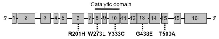 Fig. 1 Structure of human GLB1 gene and Morquio B (MBD)-related mutations.1
Key structural properties of GLB1:
- Conservative TIM barrel-shaped catalytic domain
- Stable hydrophobic core and disulfide bonds
- Two key catalytic glutamate residues
- The substrate combination of pocket accurate configuration
Functions of GLB1
The main function of the GLB1 gene is to hydrolyze the β-galactosidic bonds at the ends of glycolipids and glycoproteins. It plays a key role in a variety of physiological and pathological processes, and its functional deficiency is directly related to severe metabolic diseases.
| Function | Description |
| Glycolipid metabolism | The decomposition of substrates containing terminal β -galactose, such as GM1 ganglioside and glycoprotein oligosaccharide chains, in lysosomes is a core step in the sphingolipid metabolic pathway. |
| Maintenance of cellular homeostasis | By promptly eliminating metabolic intermediate products, the abnormal accumulation of toxic substances in lysosomes can be prevented, maintaining the normal function of cells and the autophagy cycle. |
| Pathological association of genetic diseases | The complete or partial loss of its enzymatic activity is the fundamental molecular cause leading to GM1 ganglioside storage disease and Morquio B syndrome (mucopolysaccharide storage disease type IVB). |
| Biomarker potential | Enzyme activity levels or gene mutation conditions can serve as key diagnostic indicators for related lysosomal storage disorders and targets for neonatal screening. |
| Development and nervous system support | Especially in infancy and early childhood, its normal function is crucial for the formation of myelin sheaths and the development of neurons in the central nervous system. |
Especially in infancy and early childhood, its normal function is crucial for the formation of myelin sheaths and the development of neurons in the central nervous system.
Applications of GLB1 and GLB1 Antibody in Literature
1. Sun, Jie, et al. "A Glb1-2A-mCherry reporter monitors systemic aging and predicts lifespan in middle-aged mice." Nature Communications 13.1 (2022): 7028. https://doi.org/10.1038/s41467-022-34801-9
In this study, a Glb1-2A-mCherry (GAC) senescent reporter mouse model was constructed. Its signal was linearly correlated with aging, indicating cardiac hypertrophy and shortened lifespan, and responded to senolytics drugs in the pulmonary fibrosis model. This model provides a new tool for systematic monitoring of aging and evaluation of intervention effects.
2. Yuskiv, Nataliya, Katsumi Higaki, and Sylvia Stockler-Ipsiroglu. "Morquio B disease. Disease characteristics and treatment options of a distinct GLB1-related dysostosis multiplex." International Journal of Molecular Sciences 21.23 (2020): 9121. https://doi.org/10.3390/ijms21239121
The article indicates that Morquio B disease is a lysosomal storage disorder related to the GLB1 gene, mainly manifested as abnormal bone development (pure skeletal type) or combined neuropathy (MBD plus type). The characteristics include growth retardation, spinal deformity and joint laxity. Currently, treatment directions such as cytokine targeting and gene therapy are being explored.
3. Rodriguez-Antiguedad, Jon, et al. "Genotype–Phenotype Relations for the Dystonia-Parkinsonism Genes GLB1, SLC6A3, SLC30A10, SLC39A14, and PLA2G6: MDSGene Systematic Review." International Journal of Molecular Sciences 26.9 (2025): 4074. https://doi.org/10.3390/ijms26094074
This article reviews the phenotypic and genotypic associations of dystonia Parkinson's syndrome related to five genes such as GLB1. Research has found that there are differences in the age of onset and phenotypic composition among patients with different gene diseases. Some are mainly characterized by dystonia, and the DYT naming prefix needs to be reconsidered. Early identification is crucial for treatable diseases.
4. Piloto, Ana Margarida, et al. "Plastic antibodies tailored on quantum dots for an optical detection of myoglobin down to the femtomolar range." Scientific reports 8.1 (2018): 4944. https://doi.org/10.1002/jmd2.12324
This case report presents a case of β -galactosidase deficiency caused by compound heterozygous mutations in the GLB1 gene, which is characterized by a muscle phenotype with severe proximal muscle weakness as a prominent feature, accompanied by decreased enzyme activity and increased excretion of keratin sulfate in urine.
5. Stockler‐Ipsiroglu, Sylvia, et al. "Morquio‐like dysostosis multiplex presenting with neuronopathic features is a distinct GLB1‐related phenotype." JIMD reports 60.1 (2021): 23-31. https://doi.org/10.1002/jmd2.12211
This study described 17 Brazilian patients with GLB1-related bone dysplasia and found that the majority presented with Morquio B disease (MBD) characteristics. Among them, the majority (12 cases) were complicated with neuropathy (MBD plus type), and spinal cord compression was common. Mutation analysis revealed a high frequency of the T500A allele. The research emphasized the necessity of developing new therapies for its progressive skeletal manifestations.
Creative Biolabs: GLB1 Antibodies for Research
Creative Biolabs specializes in the production of high-quality GLB1 antibodies for research and industrial applications. Our portfolio includes monoclonal antibodies tailored for ELISA, Flow Cytometry, Western blot, immunohistochemistry, and other diagnostic methodologies.
- Custom GLB1 Antibody Development: Tailor-made solutions to meet specific research requirements.
- Bulk Production: Large-scale antibody manufacturing for industry partners.
- Technical Support: Expert consultation for protocol optimization and troubleshooting.
- Aliquoting Services: Conveniently sized aliquots for long-term storage and consistent experimental outcomes.
For more details on our GLB1 antibodies, custom preparations, or technical support, contact us at email.
Reference
- Yuskiv, Nataliya, Katsumi Higaki, and Sylvia Stockler-Ipsiroglu. "Morquio B disease. Disease characteristics and treatment options of a distinct GLB1-related dysostosis multiplex." International Journal of Molecular Sciences 21.23 (2020): 9121. https://doi.org/10.3390/ijms21239121
Anti-GLB1 antibodies
 Loading...


Hot products 
-
Rabbit Anti-ABL1 (Phosphorylated Y245) Recombinant Antibody (V2-505716) (PTM-CBMAB-0465LY)

-
Mouse Anti-BrdU Recombinant Antibody (IIB5) (CBMAB-1038CQ)

-
Mouse Anti-FLI1 Recombinant Antibody (CBXF-0733) (CBMAB-F0435-CQ)

-
Mouse Anti-AFM Recombinant Antibody (V2-634159) (CBMAB-AP185LY)

-
Mouse Anti-ADGRE5 Recombinant Antibody (V2-360335) (CBMAB-C2088-CQ)

-
Rabbit Anti-ADRA1A Recombinant Antibody (V2-12532) (CBMAB-1022-CN)

-
Mouse Anti-EGR1 Recombinant Antibody (CBWJZ-100) (CBMAB-Z0289-WJ)

-
Mouse Anti-GLP1R Recombinant Antibody (4F3) (CBMAB-G0521-LY)

-
Rabbit Anti-CCL5 Recombinant Antibody (R0437) (CBMAB-R0437-CN)

-
Mouse Anti-FLT1 Recombinant Antibody (11) (CBMAB-V0154-LY)

-
Mouse Anti-BAX Recombinant Antibody (CBYY-0216) (CBMAB-0217-YY)

-
Mouse Anti-BIRC7 Recombinant Antibody (88C570) (CBMAB-L0261-YJ)

-
Mouse Anti-CCS Recombinant Antibody (CBFYC-1093) (CBMAB-C1150-FY)

-
Mouse Anti-ACO2 Recombinant Antibody (V2-179329) (CBMAB-A0627-YC)

-
Mouse Anti-CD24 Recombinant Antibody (SN3) (CBMAB-C1037-CQ)

-
Mouse Anti-Acetyl SMC3 (K105/K106) Recombinant Antibody (V2-634053) (CBMAB-AP052LY)

-
Mouse Anti-ARHGAP5 Recombinant Antibody (54/P190-B) (CBMAB-P0070-YC)

-
Mouse Anti-FOXL1 Recombinant Antibody (CBXF-0845) (CBMAB-F0462-CQ)

-
Mouse Anti-ATP1A2 Recombinant Antibody (M7-PB-E9) (CBMAB-A4013-YC)

-
Mouse Anti-FTH1 Recombinant Antibody (CBXF-1896) (CBMAB-F3426-CQ)

- AActivation
- AGAgonist
- APApoptosis
- BBlocking
- BABioassay
- BIBioimaging
- CImmunohistochemistry-Frozen Sections
- CIChromatin Immunoprecipitation
- CTCytotoxicity
- CSCostimulation
- DDepletion
- DBDot Blot
- EELISA
- ECELISA(Cap)
- EDELISA(Det)
- ESELISpot
- EMElectron Microscopy
- FFlow Cytometry
- FNFunction Assay
- GSGel Supershift
- IInhibition
- IAEnzyme Immunoassay
- ICImmunocytochemistry
- IDImmunodiffusion
- IEImmunoelectrophoresis
- IFImmunofluorescence
- IGImmunochromatography
- IHImmunohistochemistry
- IMImmunomicroscopy
- IOImmunoassay
- IPImmunoprecipitation
- ISIntracellular Staining for Flow Cytometry
- LALuminex Assay
- LFLateral Flow Immunoassay
- MMicroarray
- MCMass Cytometry/CyTOF
- MDMeDIP
- MSElectrophoretic Mobility Shift Assay
- NNeutralization
- PImmunohistologyp-Paraffin Sections
- PAPeptide Array
- PEPeptide ELISA
- PLProximity Ligation Assay
- RRadioimmunoassay
- SStimulation
- SESandwich ELISA
- SHIn situ hybridization
- TCTissue Culture
- WBWestern Blot
