HADH Antibodies
Background
The HADH gene encodes hydroxyacyl-coA dehydrogenase, which is A component of mitochondrial triple-function proteins and is mainly distributed in the mitochondrial matrix of liver and cardiomyocytes. It participates in maintaining the homeostasis of the body's energy metabolism by catalyzing the dehydrogenation reaction of hydroxyacyl-coA in the β -oxidation of fatty acids. The deficiency of this enzyme can cause the hereditary metabolic disease HADH deficiency, which is clinically manifested as hypoketogenic hypoglycemia and cardiomyopathy. The three-dimensional structure analysis of it reveals the unique tetramer conformation and substrate binding characteristics, providing an important model for the molecular diagnosis and targeted therapy research of congenital metabolic disorders, and has key scientific value for understanding the regulatory mechanism of metabolic pathways.
Structure of HADH
The mitochondrial hydroxyacyl-coA dehydrogenase encoded by the HADH gene is A tri-functional protein complex composed of approximately 34.5 kDa α subunits. This enzyme shows significant structural conservation in different species:
| Species | Human | Mouse | Rat | Bovine |
| Molecular Weight (kDa) | 34.3 | 34.1 | 34.2 | 34.4 |
| Primary Structural Differences | Highly conserved catalytic domains | 95% sequence homology | Similar substrate binding sites | Stable tetramer conformation |
This protein is composed of 308 amino acid residues, and its three-dimensional structure presents a typical α/β folding pattern. The active center contains the key glutamate-glycine-serine tripeptide structure, which stabilizes the binding of coenzyme NAD+ through a precise hydrogen bond network. The unique "Rossmann fold" conformation is responsible for coordinating substrate recognition and catalytic processes, while conserved cysteine residues are involved in maintaining the stability of the enzyme's quaternary structure. This structural feature explains its key function in the fatty acid β -oxidation pathway.
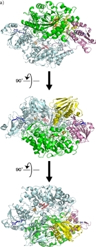 Fig. 1 Overview of recombinant HADH structure.1
Key structural properties of HADH:
- Typical α/β fold three-domain structure
- Conserved catalytic triplets constitute the active center
- Rossmann folded conformation characteristic of the NAD+ coenzyme binding domain
- Four dimer interface between salt bridge network to maintain structural stability
Functions of HADH
The core function of the protein encoded by the HADH gene is to catalyze the key step in the β -oxidation of fatty acids. This enzyme directly promotes the tricarboxylic acid cycle by specifically recognizing medium and short-chain hydroxyacyl-COA substrates and converting them into the corresponding ketoacyl-COA. Its functional system is specifically manifested as:
| Function | Description |
| Energy metabolism-driven | Dehydrogenation of 3-hydroxyl-coa is catalyzed in the mitochondrial matrix to directly generate NADH, which provides an electron source for the respiratory chain. |
| Substrate channel regulation | Form metabolic channels with other subunits in the trifunctional protein complex to ensure efficient conversion of fatty acid oxidation intermediates. |
| Maintenance of metabolic homeostasis | By regulating the decomposition rate of medium and short-chain fatty acids, abnormal accumulation of lipid intermediates in the liver and heart muscle can be prevented. |
| Regulation of ketone body formation | Effect acetyl CoA flux, indirect intrahepatic ketone body to synthesize level adjustment. |
| Associated with congenital metabolic diseases | Enzyme activity deficiency leads to HADH deficiency, which is clinically manifested as hypoketogenic hypoglycemia and metabolic crisis like Rey's syndrome. |
The catalytic kinetic characteristics of this enzyme are manifested as its specific recognition of the C4-C12 hydroxyacyl-COA substrate, and its reaction rate is regulated by the mitochondrial NAD+/NADH ratio. This regulatory mechanism ensures the energy supply switching under starvation conditions.
Applications of HADH and HADH Antibody in Literature
1. Ye, Haowen, et al. "HADH may be the target molecule of early vascular endothelial impairment in T2DM." Frontiers in Cardiovascular Medicine 9 (2022): 963916. https://doi.org/10.3389/fcvm.2022.963916
Through bioinformatics analysis, it was found that among the early vascular endothelial cell dysfunction caused by type 2 diabetes, the HADH gene has the highest diagnostic value. Experimental verification shows that HADH expression is significantly downregulated in human coronary endothelial cells treated with high glucose and palmitic acid, suggesting that it can serve as an early intervention target.
2. Shen, Congcong, et al. "Downregulation of HADH promotes gastric cancer progression via Akt signaling pathway." Oncotarget 8.44 (2017): 76279. https://doi.org/10.18632/oncotarget.19348
This study reveals for the first time that HADH inhibits tumor progression in gastric cancer by regulating the Akt signaling pathway. The expression of HADH in gastric cancer tissues decreases with the progression of the stage. Its down-regulation significantly promotes the migration and invasion of cancer cells, and this effect can be reversed by PI3K inhibitors.
3. Danquah, Bright D., et al. "Mass Spectrometric analysis of antibody—Epitope peptide complex dissociation: Theoretical concept and practical procedure of binding strength characterization." Molecules 25.20 (2020): 4776. https://doi.org/10.1109/JTEHM.2021.3104966
Studies have confirmed that HADH functional defects can disrupt the REDOX state of myocardial mitochondria. In the neonatal rat model, the REDOX ratio of the mutant heart was significantly reduced by 31.9%. This severe energy metabolism disorder is the key cause of heart failure and even sudden death.
4. Hashemian, Somayyeh, et al. "Genotyping of ABCC8, KCNJ11, and HADH in Iranian Infants with Congenital Hyperinsulinism." Case Reports in Endocrinology 2021.1 (2021): 8826174. https://doi.org/10.1155/2021/8826174
This study screened for pathogenic genes in children with congenital hyperinsulinemia. Among the 20 children patients, most of the variations were concentrated in the ABCC8 gene, and HADH is also an important pathogenic gene causing this disease, especially in the population with a high rate of consanguineous marriage, which requires special attention.
5. Çamtosun, Emine, et al. "A deep intronic HADH splicing mutation (c. 636+ 471G> T) in a congenital hyperinsulinemic hypoglycemia case: long term clinical course." Journal of clinical research in pediatric endocrinology 7.2 (2015): 144. https://doi.org/10.4274/jcrpe.1963
This study shows that the HADH gene defect is different from other fatty acid oxidation disorders. Its typical feature is hypoglycemia accompanied by hyperinsulinemia in neonates or infants. This article reports a case caused by a deep intron splicing mutation, presenting its long-term clinical course.
Creative Biolabs: HADH Antibodies for Research
Creative Biolabs specializes in the production of high-quality HADH antibodies for research and industrial applications. Our portfolio includes monoclonal antibodies tailored for ELISA, Flow Cytometry, Western blot, immunohistochemistry, and other diagnostic methodologies.
- Custom HADH Antibody Development: Tailor-made solutions to meet specific research requirements.
- Bulk Production: Large-scale antibody manufacturing for industry partners.
- Technical Support: Expert consultation for protocol optimization and troubleshooting.
- Aliquoting Services: Conveniently sized aliquots for long-term storage and consistent experimental outcomes.
For more details on our HADH antibodies, custom preparations, or technical support, contact us at email.
Reference
- Reed, Timothy, et al. "Crystal structure of histamine dehydrogenase from Nocardioides simplex." Journal of Biological Chemistry 285.33 (2010): 25782-25791. https://doi.org/10.1074/jbc.M109.084301
Anti-HADH antibodies
 Loading...


Hot products 
-
Mouse Anti-CD46 Recombinant Antibody (CBFYC-0076) (CBMAB-C0085-FY)

-
Mouse Anti-ENPP1 Recombinant Antibody (CBFYE-0159) (CBMAB-E0375-FY)

-
Rat Anti-(1-5)-α-L-Arabinan Recombinant Antibody (V2-501861) (CBMAB-XB0003-YC)

-
Mouse Anti-CARD11 Recombinant Antibody (CBFYC-0811) (CBMAB-C0866-FY)

-
Mouse Anti-4-Hydroxynonenal Recombinant Antibody (V2-502280) (CBMAB-C1055-CN)

-
Mouse Anti-CD1C Recombinant Antibody (L161) (CBMAB-C2173-CQ)

-
Mouse Anti-GFAP Recombinant Antibody (20) (CBMAB-G2914-LY)

-
Mouse Anti-BLNK Recombinant Antibody (CBYY-0623) (CBMAB-0626-YY)

-
Mouse Anti-BHMT Recombinant Antibody (CBYY-0547) (CBMAB-0550-YY)

-
Mouse Anti-ASB9 Recombinant Antibody (1D8) (CBMAB-A0529-LY)

-
Mouse Anti-BCL6 Recombinant Antibody (CBYY-0442) (CBMAB-0445-YY)

-
Mouse Anti-BANF1 Recombinant Antibody (3F10-4G12) (CBMAB-A0707-LY)

-
Mouse Anti-CD24 Recombinant Antibody (2Q1282) (CBMAB-C1624-CN)

-
Mouse Anti-ARID3A Antibody (A4) (CBMAB-0128-YC)

-
Rabbit Anti-BAD (Phospho-Ser136) Recombinant Antibody (CAP219) (CBMAB-AP536LY)

-
Mouse Anti-CAT Recombinant Antibody (724810) (CBMAB-C8431-LY)

-
Rabbit Anti-CBL Recombinant Antibody (D4E10) (CBMAB-CP0149-LY)

-
Mouse Anti-AAV-5 Recombinant Antibody (V2-503417) (CBMAB-V208-1369-FY)

-
Rat Anti-CCR2 Recombinant Antibody (475301) (CBMAB-C1338-LY)

-
Mouse Anti-BIRC3 Recombinant Antibody (315304) (CBMAB-1214-CN)

- AActivation
- AGAgonist
- APApoptosis
- BBlocking
- BABioassay
- BIBioimaging
- CImmunohistochemistry-Frozen Sections
- CIChromatin Immunoprecipitation
- CTCytotoxicity
- CSCostimulation
- DDepletion
- DBDot Blot
- EELISA
- ECELISA(Cap)
- EDELISA(Det)
- ESELISpot
- EMElectron Microscopy
- FFlow Cytometry
- FNFunction Assay
- GSGel Supershift
- IInhibition
- IAEnzyme Immunoassay
- ICImmunocytochemistry
- IDImmunodiffusion
- IEImmunoelectrophoresis
- IFImmunofluorescence
- IGImmunochromatography
- IHImmunohistochemistry
- IMImmunomicroscopy
- IOImmunoassay
- IPImmunoprecipitation
- ISIntracellular Staining for Flow Cytometry
- LALuminex Assay
- LFLateral Flow Immunoassay
- MMicroarray
- MCMass Cytometry/CyTOF
- MDMeDIP
- MSElectrophoretic Mobility Shift Assay
- NNeutralization
- PImmunohistologyp-Paraffin Sections
- PAPeptide Array
- PEPeptide ELISA
- PLProximity Ligation Assay
- RRadioimmunoassay
- SStimulation
- SESandwich ELISA
- SHIn situ hybridization
- TCTissue Culture
- WBWestern Blot
