ID2 Antibodies
Background
The ID2 gene encodes a helical-ring-helical transcription factor, which is mainly present in the nervous system, immune organs and skeletal muscle development of vertebrates. This protein regulates the cell differentiation cycle and maintains the characteristics of stem cells by forming heterodimers to inhibit the activity of other transcription factors. During the embryonic development stage, the ID2 gene plays a crucial role in the migration of neural crest cells and the formation of lymphoid organs. An imbalance in its expression will directly affect the process of tissue differentiation. This gene was first identified by Robert Benezra's team in 1991. Subsequent studies have revealed that it participates in the tumor suppression pathway by regulating cyclin-dependent stimulator inhibitors, providing an important molecular target for cancer biology. Its multifunctional regulatory mechanism continuously drives the research progress in the fields of developmental biology and tumor treatment.
Structure of ID2
Myoglobin is a relatively small protein with a molecular weight of approximately 16.7 kDa. This weight may slightly vary between species due to minor differences in amino acid sequence.
| Species | Human | Mouse | Rat | Bovine |
| Molecular Weight (kDa) | 14.5 | 14.3 | 14.4 | 14.6 |
| Primary Structural Differences | Contains four conservative domain structure | N-terminal sequence is slightly different | Highly conserved DNA binding domain | With the human homology of 95% |
The ID2 protein is composed of 134 amino acids, and its core structural feature is a conserved HLH domain, which consists of two α -helicles and an interventional ring region. This HLH domain enables ID2 to form heterodimers with other bHLH transcription factors. However, due to the incomplete function of the positively charged basic region in its DNA-binding domain, the formed complex cannot effectively bind to DNA, thereby exerting its dominant negative regulatory effect. This unique molecular mechanism is the structural basis for ID2 as a key inhibitor of cell differentiation.
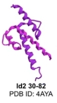 Fig. 1 Structures of the homodimers of the fragments Id2 30–82.1
Key structural properties of ID2:
- Helix-loop-helix (HLH) dimerization domain
- Dysfunctional DNA-binding regions
- Conserved N-terminal region and easily degraded C-terminal sequence
Functions of ID2
The core function of the ID2 gene is to regulate cell differentiation and proliferation. However, it is also widely involved in key physiological and pathological processes such as developmental programming, immune response and tumorigenesis.
| Function | Description |
| Differentiation inhibition | By inhibiting the tissue-specific bHLH transcription factor, the terminal differentiation process of various cell lineages (such as neural, muscular, and immune) is blocked. |
| Cell cycle promotion | Maintain cells in a proliferative state and directly regulate the expression of cyclin-dependent kinase inhibitors. |
| Regulation of embryonic development | In the lymph organ formation and migration of neural crest, angiogenesis and play a key role in the process of embryonic development. |
| Immune regulation | Regulate the differentiation and function of lymphocytes and influence the balance between innate immunity and adaptive immunity. |
| Tumorigenesis participation | In a variety of abnormally high expression in cancer, inhibit differentiation, promoting the transformation of epithelial - stromal tumor progression. |
The heterodimer formed by ID2 and bHLH transcription factors such as E protein cannot effectively bind to DNA. This "dominant negative regulation" mechanism makes it play the role of a molecular switch in cell fate determination, and its activity level directly determines whether the cell enters the differentiation process or maintains the proliferation state.
Applications of ID2 and ID2 Antibody in Literature
1. Li, Yiming, et al. "Id2 epigenetically controls CD8+ T-cell exhaustion by disrupting the assembly of the Tcf3-LSD1 complex." Cellular & Molecular Immunology 21.3 (2024): 292-308. https://doi.org/10.1038/s41423-023-01118-6
The article indicates that ID2 interferes with the formation of the TCF3-TAL1 complex through its HLH domain, preventing Tcf3 from binding to the demethylase LSD1, thereby enhancing chromatin accessibility in the Slamf6 promoter region and promoting the generation of stem cell-like exhausted T cells. Targeting LSD1 can reverse the anti-tumor functional defect caused by ID2 deficiency.
2. Machold, Robert, et al. "Id2 GABAergic interneurons comprise a neglected fourth major group of cortical inhibitory cells." elife 12 (2023): e85893. https://doi.org/10.7554/eLife.85893
Research has confirmed that Id2 can serve as a key genetic marker for specifically identifying and studying a particular type of inhibitory neurons in the cerebral cortex. These ID2-positive neurons are mainly glial morphoblasts (NGFCs), which possess unique electrophysiological characteristics and exhibit specific activity patterns during sleep. This discovery provides an important genetic tool for in-depth exploration of such cells.
3. Vázquez-Cabrera, Guillermo, et al. "ID2-ETS2 axis regulates the transcriptional acquisition of pro-tumoral microglia phenotype in glioma." Cell Death & Disease 15.7 (2024): 512. https://doi.org/10.1038/s41419-024-06903-3
Studies have confirmed that in glioblastoma, tumor cells up-regulate the expression of ID2, enabling it to interact with the transcription factor ETS2, thereby driving microglia to transform into a phenotype that supports tumor growth. The ID2-ETS2 transcriptional axis is a key mechanism for regulating the function of tumor-associated microglia, providing a new target for potential therapeutic interventions.
4. Mao, Weipu, et al. "ID2 inhibits bladder cancer progression and metastasis via PI3K/AKT signaling pathway." Frontiers in Cell and Developmental Biology 9 (2021): 738364. https://doi.org/10.3389/fcell.2021.738364
Studies have confirmed that in bladder cancer, the expression of ID2 is significantly down-regulated, and its high level indicates a better prognosis. Functionally, ID2 effectively inhibits the proliferation, migration and invasion of tumor cells by suppressing the PI3K/AKT signaling pathway, thereby exerting an anti-cancer effect. ID2 can serve as a potential biomarker for the progression of bladder cancer.
5. Deng, Zhongming, et al. "ID2 promotes tumor progression and metastasis in thyroid cancer." Endocrine 84.3 (2024): 1051-1063. https://doi.org/10.1007/s12020-023-03674-3
Studies have confirmed that ID2 significantly promotes the proliferation, migration, epithelial-mesenchymal transition and stem cell characteristics of tumor cells in thyroid cancer by activating the PI3K/Akt signaling pathway. Its overexpression is associated with a poor prognosis of cancer and may become a potential biomarker for enhancing the efficacy of chemotherapy and immunotherapy.
Creative Biolabs: ID2 Antibodies for Research
Creative Biolabs specializes in the production of high-quality ID2 antibodies for research and industrial applications. Our portfolio includes monoclonal antibodies tailored for ELISA, Flow Cytometry, Western blot, immunohistochemistry, and other diagnostic methodologies.
- Custom ID2 Antibody Development: Tailor-made solutions to meet specific research requirements.
- Bulk Production: Large-scale antibody manufacturing for industry partners.
- Technical Support: Expert consultation for protocol optimization and troubleshooting.
- Aliquoting Services: Conveniently sized aliquots for long-term storage and consistent experimental outcomes.
For more details on our ID2 antibodies, custom preparations, or technical support, contact us at email.
Reference
- Roschger, Cornelia, and Chiara Cabrele. "The Id-protein family in developmental and cancer-associated pathways." Cell Communication and Signaling 15.1 (2017): 7. https://doi.org/10.1186/s12964-016-0161-y
Anti-ID2 antibodies
 Loading...


Hot products 
-
Mouse Anti-COL1A2 Recombinant Antibody (CF108) (V2LY-1206-LY626)

-
Mouse Anti-GGT1 Recombinant Antibody (1F9) (CBMAB-G3273-LY)

-
Rabbit Anti-CBL Recombinant Antibody (D4E10) (CBMAB-CP0149-LY)

-
Mouse Anti-CD33 Recombinant Antibody (6C5/2) (CBMAB-C8126-LY)

-
Mouse Anti-GFAP Recombinant Antibody (24) (CBMAB-G2927-LY)

-
Mouse Anti-2C TCR Recombinant Antibody (V2-1556) (CBMAB-0951-LY)

-
Mouse Anti-BLNK Recombinant Antibody (CBYY-0623) (CBMAB-0626-YY)

-
Mouse Anti-CARTPT Recombinant Antibody (113612) (CBMAB-C2450-LY)

-
Rat Anti-C5AR1 Recombinant Antibody (8D6) (CBMAB-C9139-LY)

-
Mouse Anti-CCDC25 Recombinant Antibody (CBLC132-LY) (CBMAB-C9786-LY)

-
Mouse Anti-CCDC6 Recombinant Antibody (CBXC-0106) (CBMAB-C5397-CQ)

-
Mouse Anti-APOE Recombinant Antibody (A1) (CBMAB-0078CQ)

-
Mouse Anti-BIRC3 Recombinant Antibody (315304) (CBMAB-1214-CN)

-
Mouse Anti-AAV9 Recombinant Antibody (V2-634029) (CBMAB-AP023LY)

-
Mouse Anti-AAV8 Recombinant Antibody (V2-634028) (CBMAB-AP022LY)

-
Mouse Anti-ADRB2 Recombinant Antibody (V2-180026) (CBMAB-A1420-YC)

-
Human Anti-SARS-CoV-2 Spike Recombinant Antibody (CR3022) (CBMAB-CR014LY)

-
Mouse Anti-CCND2 Recombinant Antibody (DCS-3) (CBMAB-G1318-LY)

-
Mouse Anti-GFAP Recombinant Antibody (20) (CBMAB-G2914-LY)

-
Rat Anti-(1-5)-α-L-Arabinan Recombinant Antibody (V2-501861) (CBMAB-XB0003-YC)

- AActivation
- AGAgonist
- APApoptosis
- BBlocking
- BABioassay
- BIBioimaging
- CImmunohistochemistry-Frozen Sections
- CIChromatin Immunoprecipitation
- CTCytotoxicity
- CSCostimulation
- DDepletion
- DBDot Blot
- EELISA
- ECELISA(Cap)
- EDELISA(Det)
- ESELISpot
- EMElectron Microscopy
- FFlow Cytometry
- FNFunction Assay
- GSGel Supershift
- IInhibition
- IAEnzyme Immunoassay
- ICImmunocytochemistry
- IDImmunodiffusion
- IEImmunoelectrophoresis
- IFImmunofluorescence
- IGImmunochromatography
- IHImmunohistochemistry
- IMImmunomicroscopy
- IOImmunoassay
- IPImmunoprecipitation
- ISIntracellular Staining for Flow Cytometry
- LALuminex Assay
- LFLateral Flow Immunoassay
- MMicroarray
- MCMass Cytometry/CyTOF
- MDMeDIP
- MSElectrophoretic Mobility Shift Assay
- NNeutralization
- PImmunohistologyp-Paraffin Sections
- PAPeptide Array
- PEPeptide ELISA
- PLProximity Ligation Assay
- RRadioimmunoassay
- SStimulation
- SESandwich ELISA
- SHIn situ hybridization
- TCTissue Culture
- WBWestern Blot
