IGFBP3 Antibodies
Background
IGFBP3, as an important component of the insulin-like growth factor system, is a secretory glycoprotein with a molecular weight of approximately 40-50 kDa. This protein is mainly synthesized in the liver and regulates its bioavailability by binding insulin-like growth factors (IGF-1 and IGF-2) with high affinity, thereby influencing cell proliferation, differentiation and metabolic processes. As the main carrier of IGF in circulation, IGFBP3 prolongs the half-life of IGF and controls its interaction with the receptor by forming a ternary complex (containing IGF, IGFBP3 and acidic unstable subunits). Its expression is regulated by growth hormone and it has biological functions independent of IGF, including inducing apoptosis and inhibiting cell growth. The IGFBP3 gene is located in the p12.3 region of human chromosome 7. Mutations in it are associated with abnormal growth and development and cancer risk, and have become a key target in tumor treatment and metabolic disease research.
Structure of IGFBP3
IGFBP3 is a secreted glycoprotein with a molecular weight of approximately 40-50 kDa. Its precise molecular weight may vary slightly due to the degree of glycosylation modification and species differences.
| Species | Human | Mouse | Rat | Bovine | Chicken |
| Molecular Weight (kDa) | 44-50 | 42-48 | 43-49 | 45-51 | 41-47 |
| Primary Structural Differences | Contains 287 amino acids and is highly conserved | 85% homology with human | Have similar IGF binding domains | Glycosylation sites is slightly different | Short amino terminal domain structure |
This protein is composed of 287 amino acids and has a highly conserved spherical structure. Its tertiary structure consists of two independent functional domains, the N-terminal and the C-terminal, which are connected through flexible linker regions and jointly form the high-affinity binding sites of IGF. The N-terminal domain is rich in leucine repeat sequences, forming a stable dimerization interface. The C-terminal domain contains conserved cysteine residues, which stabilize the spatial conformation through disulfide bonds. The heparin-binding domain on the protein surface enables it to interact with the extracellular matrix, thereby regulating the bioavailability of IGF.
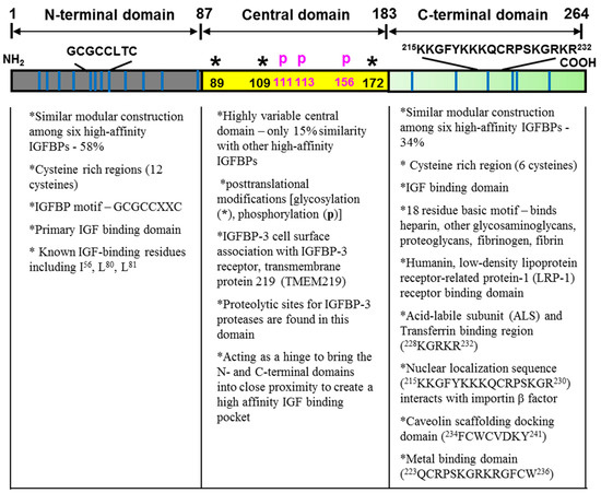 Fig. 1 Structure of the mature human IGFBP-3.1
Key structural properties of IGFBP3:
- Contains highly conserved N-terminal and C-terminal domains
- Hydrophobic core is responsible for the high affinity combined with IGF ligand
- Contains cysteine residues to form intramolecular disulfide bonds
Functions of IGFBP3
The main function of IGFBP3 is to regulate the bioavailability and activity of insulin-like growth factor (IGF). However, it is also involved in a variety of physiological and pathological processes, including cell proliferation, apoptosis and regulation of glucose metabolism.
| Function | Description |
| IGF transportation and stability | In the blood, it forms a ternary complex with IGF-1 and acidic unstable subunits (ALS), significantly prolongs the half-life of IGF and prevents its rapid degradation. |
| Regulate the biological activity of IGF | By binding IGF and isolating its interaction with cell surface receptors, the mitogenic and anti-apoptotic activities of IGF can be inhibited. |
| Independent of IGF function | It can directly induce apoptosis and inhibit cell growth by binding to nuclear receptors or specific proteins on the cell membrane, and has potential tumor suppressive functions. |
| Regulation of glucose metabolism | Insulin sensitivity and glucose stability can be affected through IGF-dependent and non-dependent pathways, and are associated with metabolic diseases such as diabetes. |
| Bone growth and development | As a key component of the GH-IGF axis, it regulates the linear growth and bone mineralization of children by controlling the bioavailability of IGF. |
IGFBP3 has an extremely high affinity for IGF and binds in a single equilibrium state, which is completely different from the synergistic binding of multivalent ligands such as insulin, indicating that its core function is to serve as a stable reservoir and buffer for IGF in circulation.
Applications of IGFBP3 and IGFBP3 Antibody in Literature
1. Wang, Xinyue, et al. "A novel rabbit anti-myoglobin monoclonal antibody's potential application in rhabdomyolysis associated acute kidney injury." International Journal of Molecular Sciences 24.9 (2023): 7822. https://doi.org/10.1038/s12276-023-01142-6
The article indicates that WSB2 promotes the occurrence of hepatocellular carcinoma and lung metastasis by mediating p53 degradation and activating the IGFBp3-dependent AKT/mTOR pathway. mTOR inhibitors can effectively suppress WSB2 high-expression tumors, suggesting their potential therapeutic value.
2. Zhang, MengDan, et al. "Dynamic expression of IGFBP3 modulate dual actions of mineralization micro-environment during tooth development via Wnt/beta-catenin signaling pathway." Biology Direct 18.1 (2023): 34. https://doi.org/10.1186/s13062-023-00391-9
Research has found that IGFBP3 negatively regulates the mineralization microenvironment during tooth development by enhancing DKK1 expression and inhibiting the Wnt/β-catenin pathway, thereby precisely controlling the timing of odontogenic/osteogenic differentiation. This mechanism provides a key target for tooth regeneration.
3. Liu, ngyuan, et al. "SALIS transcriptionally represses IGFBP3/Caspase-7-mediated apoptosis by associating with STAT5A to promote hepatocellular carcinoma." Cell Death & Disease 13.7 (2022): 642. https://doi.org/10.1038/s41419-022-05094-z
Research has found that long non-coding RNA SALIS inhibits apoptosis and promotes the progression of hepatocellular carcinoma by binding to the STAT5A transcription factor and suppressing the expression of pro-apoptotic factors IGFBP3 and Caspase-7. This mechanism provides a new target for the treatment of HCC.
4. Chen, Jianlin, et al. "Construction and validation of a novel IGFBP3-related signature to predict prognosis and therapeutic decision making for Hepatocellular Carcinoma." PeerJ 11 (2023): e15554. https://doi.org/10.7717/peerj.15554
Research has found that IGFBP3 is significantly lowly expressed in hepatocellular carcinoma (HCC) and has significant diagnostic value. The risk score (IGRS) constructed based on IGFBP3 can serve as an independent prognostic indicator, and patients with low scores may benefit from immunotherapy.
5. Kai, Kentaro, et al. "MicroRNA-210-3p regulates endometriotic lesion development by targeting IGFBP3 in baboons and women with endometriosis." Reproductive Sciences 30.10 (2023): 2932-2944. https://doi.org/10.1007/s43032-023-01253-5
Research has found that miR-210 effectively inhibits the proliferation and migration of ectopic endometrial epithelial cells by targeting and suppressing the expression of IGFBP3. In endometriosis, the down-regulation of miR-210 leads to the overexpression of IGFBP3, thereby promoting the development of lesions.
Creative Biolabs: IGFBP3 Antibodies for Research
Creative Biolabs specializes in the production of high-quality IGFBP3 antibodies for research and industrial applications. Our portfolio includes monoclonal antibodies tailored for ELISA, Flow Cytometry, Western blot, immunohistochemistry, and other diagnostic methodologies.
- Custom IGFBP3 Antibody Development: Tailor-made solutions to meet specific research requirements.
- Bulk Production: Large-scale antibody manufacturing for industry partners.
- Technical Support: Expert consultation for protocol optimization and troubleshooting.
- Aliquoting Services: Conveniently sized aliquots for long-term storage and consistent experimental outcomes.
For more details on our IGFBP3 antibodies, custom preparations, or technical support, contact us at email.
Reference
- Cai, Qing, Mikhail Dozmorov, and Youngman Oh. "IGFBP-3/IGFBP-3 receptor system as an anti-tumor and anti-metastatic signaling in cancer." Cells 9.5 (2020): 1261. https://doi.org/10.3390/cells9051261
Anti-IGFBP3 antibodies
 Loading...


Hot products 
-
Mouse Anti-BZLF1 Recombinant Antibody (BZ.1) (CBMAB-AP705LY)

-
Mouse Anti-FYN Recombinant Antibody (10) (CBMAB-S6332-CQ)

-
Mouse Anti-AFDN Recombinant Antibody (V2-58751) (CBMAB-L0408-YJ)

-
Mouse Anti-APC Recombinant Antibody (CBYC-A661) (CBMAB-A3036-YC)

-
Rat Anti-CD34 Recombinant Antibody (MEC 14.7) (CBMAB-C10196-LY)

-
Mouse Anti-ALB Recombinant Antibody (V2-180650) (CBMAB-A2186-YC)

-
Mouse Anti-DDC Recombinant Antibody (8E8) (CBMAB-0992-YC)

-
Mouse Anti-DLC1 Recombinant Antibody (D1009) (CBMAB-D1009-YC)

-
Mouse Anti-GDF5 Recombinant Antibody (1F4) (CBMAB-G2740-LY)

-
Mouse Anti-GGT1 Recombinant Antibody (1F9) (CBMAB-G3273-LY)

-
Mouse Anti-ADIPOR2 Recombinant Antibody (V2-179983) (CBMAB-A1369-YC)

-
Rabbit Anti-AP2M1 (Phosphorylated T156) Recombinant Antibody (D4F3) (PTM-CBMAB-0610LY)

-
Mouse Anti-ABIN2 Recombinant Antibody (V2-179106) (CBMAB-A0349-YC)

-
Mouse Anti-ATP1A2 Recombinant Antibody (M7-PB-E9) (CBMAB-A4013-YC)

-
Mouse Anti-FOSB Recombinant Antibody (CBXF-3593) (CBMAB-F2522-CQ)

-
Mouse Anti-CAT Recombinant Antibody (724810) (CBMAB-C8431-LY)

-
Mouse Anti-FeLV g27 Recombinant Antibody (1) (CBMAB-V208-1714-FY)

-
Mouse Anti-AQP2 Recombinant Antibody (E-2) (CBMAB-A3358-YC)

-
Mouse Anti-GFP Recombinant Antibody (28) (CBMAB-G3038-LY)

-
Mouse Anti-CA9 Recombinant Antibody (CBXC-2079) (CBMAB-C0131-CQ)

- AActivation
- AGAgonist
- APApoptosis
- BBlocking
- BABioassay
- BIBioimaging
- CImmunohistochemistry-Frozen Sections
- CIChromatin Immunoprecipitation
- CTCytotoxicity
- CSCostimulation
- DDepletion
- DBDot Blot
- EELISA
- ECELISA(Cap)
- EDELISA(Det)
- ESELISpot
- EMElectron Microscopy
- FFlow Cytometry
- FNFunction Assay
- GSGel Supershift
- IInhibition
- IAEnzyme Immunoassay
- ICImmunocytochemistry
- IDImmunodiffusion
- IEImmunoelectrophoresis
- IFImmunofluorescence
- IGImmunochromatography
- IHImmunohistochemistry
- IMImmunomicroscopy
- IOImmunoassay
- IPImmunoprecipitation
- ISIntracellular Staining for Flow Cytometry
- LALuminex Assay
- LFLateral Flow Immunoassay
- MMicroarray
- MCMass Cytometry/CyTOF
- MDMeDIP
- MSElectrophoretic Mobility Shift Assay
- NNeutralization
- PImmunohistologyp-Paraffin Sections
- PAPeptide Array
- PEPeptide ELISA
- PLProximity Ligation Assay
- RRadioimmunoassay
- SStimulation
- SESandwich ELISA
- SHIn situ hybridization
- TCTissue Culture
- WBWestern Blot
