IL1RAP Antibodies
Background
The IL1RAP gene encodes the interleukin-1 receptor accessory protein, which is an important transmembrane protein mainly expressed on the surface of immune cells and certain tumor cells. This protein acts as a key co-receptor of the IL-1 receptor family and participates in mediating the signal transduction of cytokines such as IL-1 and IL-33, thereby regulating inflammatory responses, immune responses, and hematopoietic processes. Studies have found that IL1RAP is often overexpressed in hematological tumors such as acute myeloid leukemia. It promotes the proliferation and survival of cancer cells by activating downstream NF-κB and MAPK signaling pathways, making it regarded as a potential immunotherapy target. In recent years, monoclonal antibodies targeting IL1RAP and CAR-T therapies have shown anti-tumor effects in preclinical studies, providing a new direction for targeted intervention in related diseases.
Structure of IL1RAP
The IL1RAP gene encodes an important transmembrane protein with a molecular weight of approximately 80-85 kDa. This value may vary slightly in different cell types depending on the different splicing variants and glycosylation modification states. The following table lists the key characteristics of IL1RAP proteins from different sources:
| Species | Human | Mouse | Rat |
| Molecular Weight (kDa) | Approximately 85 | Approximately 82 | Approximately 83 |
| Primary Structural Differences | The common receptor for IL-1/IL-33 signaling, a target for cancer treatment | Involved in inflammation and immune regulation, highly homologous to human functions | Often used in the study of neural inflammation and immune related diseases |
The IL1RAP protein contains multiple domains. Its core extracellular region is composed of three immunoglobulin-like domains, which are mainly responsible for binding to IL-1 receptors (such as IL1R1) and cytokines. Its transmembrane region and intracellular tail region do not have kinase activity, but through interaction with adaptor proteins such as MyD88, they are crucial for the activation of downstream NF-κB and MAPK signaling pathways. This structural feature makes it a key molecule in regulating innate immune responses and inflammatory processes.
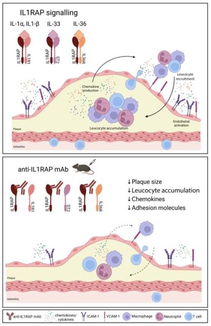 Fig. 1 IL1RAP Blockade: A Novel Therapeutic Strategy to Reduce Atherosclerotic Plaque Burden and Inflammation.1
Key structural properties of IL1RAP:
- Transmembrane co-receptor structure
- Extracellular domain-mediated protein interaction
- Intracellular domain lacks kinase activity
Functions of IL1RAP
The core function of the IL1RAP protein is to act as a necessary co-receptor of the interleukin-1 receptor family. It not only regulates inflammatory responses but also participates in a variety of important pathological and physiological processes.
| Function | Description |
| Inflammatory Signal Transduction | It forms functional receptor complexes by binding to IL-1R1 or IL-33R, initiating MyD88-dependent NF-κB and MAPK signaling pathways, which are key nodes in the innate immune response. |
| Immune Cell Activation | It is expressed on the surface of immune cells such as mast cells and macrophages, mediating the activation, proliferation and cytokine release triggered by IL-1 and IL-33. |
| Tumor Promotion | It is highly expressed in various leukemia and solid tumors. By continuously activating pro-survival signaling pathways, it supports the proliferation, resistance, and immune evasion of cancer cells. |
| Hematopoietic Regulation | It functions in hematopoietic stem cells and progenitor cells, influencing their proliferation and differentiation, and is related to the occurrence and development of hematological diseases such as myeloproliferative disorders. |
| Neuroinflammation involvement | Expressed in central nervous immune cells such as microglia, it participates in the neuroinflammatory responses in neurodegenerative diseases and brain injuries. |
The signal transduction of IL1RAP exhibits a "dual-effect switch" characteristic: On one hand, its intracellular domain has no catalytic activity on its own and relies entirely on adaptor proteins to transmit signals; on the other hand, its expression level and subcellular localization strictly regulate the intensity and specificity of IL-1 family signaling, making it an important intervention target in the treatment of inflammation and tumors.
Applications of IL1RAP and IL1RAP Antibody in Literature
1. Mulholland, Megan, et al. "Interleukin-1 receptor accessory protein blockade limits the development of atherosclerosis and reduces plaque inflammation." Cardiovascular Research 120.6 (2024): 581-595. https://doi.org/10.1093/cvr/cvae046
Studies have shown that the use of novel antibodies to block IL1RAP can significantly reduce the burden of atherosclerotic plaques and inflammation in mice, and decrease the expression of factors related to white blood cell recruitment. IL1RAP is a key co-receptor in the IL-1, IL-33, and IL-36 signaling pathways, and its inhibition provides a new target for the treatment of atherosclerosis.
2. Métois, Arnaud, et al. "IL1RAP is an immunotherapeutic target for normal karyotype triple-mutated acute myeloid leukemia." Biomarker Research 13.1 (2025): 61. https://doi.org/10.1186/s40364-025-00769-z
This study found that IL1RAP is highly specifically expressed on the surface of NKt-AML leukemia cells, and is associated with poor prognosis and transplant resistance in patients. The IL1RAP antibody can induce its internalization, suggesting its potential as a target for antibody-drug conjugates in treatment.
3. Badi, Yusef Eamon, et al. "IL1RAP expression and the enrichment of IL‐33 activation signatures in severe neutrophilic asthma." Allergy 78.1 (2023): 156-167. https://doi.org/10.1111/all.15487
The study found that the gene signatures activated by IL-33 were significantly enriched in the sputum of patients with neutrophilic and mixed granulocytic asthma, which was associated with the high expression of the IL1RAP co-receptor, suggesting that anti-IL-33 therapy should be extended to the T2 low-type asthma population.
4. Zhang, Yang, et al. "Innate immune mediator, Interleukin-1 receptor accessory protein (IL1RAP), is expressed and pro-tumorigenic in pancreatic cancer." Journal of Hematology & Oncology 15.1 (2022): 70. https://doi.org/10.1186/s13045-022-01286-4
The study found that IL1RAP is widely expressed in pancreatic cancer and its high expression is associated with poor prognosis. Knockdown of IL1RAP can significantly inhibit the growth and invasion ability of cancer cells, and the downstream IRAK4 inhibitor is effective in slowing tumor progression in preclinical models.
5. Zettergren, Anna, et al. "Association of IL 1 RAP-related genetic variation with cerebrospinal fluid concentration of Alzheimer-associated tau protein." Scientific reports 9.1 (2019): 2460. https://doi.org/10.1038/s41598-018-36650-3
The study found that the IL1RAP gene variation has no direct correlation with the risk of Alzheimer's disease, but specific SNPs (such as rs9877502) are significantly associated with the level of tau protein in the cerebrospinal fluid of patients, suggesting that this gene may affect the intensity of disease progression.
Creative Biolabs: IL1RAP Antibodies for Research
Creative Biolabs specializes in the production of high-quality IL1RAP antibodies for research and industrial applications. Our portfolio includes monoclonal antibodies tailored for ELISA, Flow Cytometry, Western blot, immunohistochemistry, and other diagnostic methodologies.
- Custom IL1RAP Antibody Development: Tailor-made solutions to meet specific research requirements.
- Bulk Production: Large-scale antibody manufacturing for industry partners.
- Technical Support: Expert consultation for protocol optimization and troubleshooting.
- Aliquoting Services: Conveniently sized aliquots for long-term storage and consistent experimental outcomes.
For more details on our IL1RAP antibodies, custom preparations, or technical support, contact us at email.
Reference
- Mulholland, Megan, et al. "Interleukin-1 receptor accessory protein blockade limits the development of atherosclerosis and reduces plaque inflammation." Cardiovascular Research 120.6 (2024): 581-595. Distributed under Open Access license CC BY 4.0, without modification. https://doi.org/10.1093/cvr/cvae046
Anti-IL1RAP antibodies
 Loading...


Hot products 
-
Mouse Anti-CRTAM Recombinant Antibody (CBFYC-2235) (CBMAB-C2305-FY)

-
Mouse Anti-AKT1 Recombinant Antibody (V2-180546) (CBMAB-A2070-YC)

-
Rat Anti-CD63 Recombinant Antibody (7G4.2E8) (CBMAB-C8725-LY)

-
Mouse Anti-ARG1 Recombinant Antibody (CBYCL-103) (CBMAB-L0004-YC)

-
Mouse Anti-CEMIP Recombinant Antibody (3C12) (CBMAB-K0296-LY)

-
Mouse Anti-ARID1B Recombinant Antibody (KMN1) (CBMAB-A3546-YC)

-
Mouse Anti-BIRC5 Recombinant Antibody (6E4) (CBMAB-CP2646-LY)

-
Mouse Anti-DLL4 Recombinant Antibody (D1090) (CBMAB-D1090-YC)

-
Mouse Anti-GFAP Recombinant Antibody (24) (CBMAB-G2927-LY)

-
Mouse Anti-COL12A1 Recombinant Antibody (CBYY-C3117) (CBMAB-C4560-YY)

-
Mouse Anti-ATP1B3 Recombinant Antibody (1E9) (CBMAB-A4021-YC)

-
Rabbit Anti-B2M Recombinant Antibody (CBYY-0059) (CBMAB-0059-YY)

-
Mouse Anti-ACLY Recombinant Antibody (V2-179314) (CBMAB-A0610-YC)

-
Rabbit Anti-DLK1 Recombinant Antibody (9D8) (CBMAB-D1061-YC)

-
Mouse Anti-BIRC3 Recombinant Antibody (315304) (CBMAB-1214-CN)

-
Rabbit Anti-AKT2 (Phosphorylated S474) Recombinant Antibody (V2-556130) (PTM-CBMAB-0605LY)

-
Mouse Anti-CD63 Recombinant Antibody (CBXC-1200) (CBMAB-C1467-CQ)

-
Mouse Anti-BSN Recombinant Antibody (219E1) (CBMAB-1228-CN)

-
Rat Anti-CD34 Recombinant Antibody (MEC 14.7) (CBMAB-C10196-LY)

-
Mouse Anti-APP Recombinant Antibody (DE2B4) (CBMAB-1122-CN)

- AActivation
- AGAgonist
- APApoptosis
- BBlocking
- BABioassay
- BIBioimaging
- CImmunohistochemistry-Frozen Sections
- CIChromatin Immunoprecipitation
- CTCytotoxicity
- CSCostimulation
- DDepletion
- DBDot Blot
- EELISA
- ECELISA(Cap)
- EDELISA(Det)
- ESELISpot
- EMElectron Microscopy
- FFlow Cytometry
- FNFunction Assay
- GSGel Supershift
- IInhibition
- IAEnzyme Immunoassay
- ICImmunocytochemistry
- IDImmunodiffusion
- IEImmunoelectrophoresis
- IFImmunofluorescence
- IGImmunochromatography
- IHImmunohistochemistry
- IMImmunomicroscopy
- IOImmunoassay
- IPImmunoprecipitation
- ISIntracellular Staining for Flow Cytometry
- LALuminex Assay
- LFLateral Flow Immunoassay
- MMicroarray
- MCMass Cytometry/CyTOF
- MDMeDIP
- MSElectrophoretic Mobility Shift Assay
- NNeutralization
- PImmunohistologyp-Paraffin Sections
- PAPeptide Array
- PEPeptide ELISA
- PLProximity Ligation Assay
- RRadioimmunoassay
- SStimulation
- SESandwich ELISA
- SHIn situ hybridization
- TCTissue Culture
- WBWestern Blot
