MSH3 Antibodies
Background
The MSH3 gene encodes a DNA mismatch repair protein, which mainly exists in the cell nucleus of eukaryotes. This protein recognizes and binds to the insertion/deletion loops during DNA replication by forming the MutSβ complex, initiating the DNA damage repair pathway to maintain genomic stability. In patients with hereditary colorectal cancer, MSH3 gene mutations often lead to increased microsatellite instability and defects in mismatch repair function. This gene was jointly identified by multiple research teams in 1996. The study of its function has provided an important basis for the molecular diagnosis of hereditary tumors such as Lynch syndrome. As a key component of the DNA repair system, the study of the mechanism of action of MSH3 protein has deepened our understanding of maintaining genomic stability, regulating the cell cycle, and tumorigenesis and development.
Structure of MSH3
MSH3 is a medium-sized protein with a molecular weight of approximately 110 kDa. This protein is encoded by the 5q14 region of the human chromosome and contains 1,137 amino acid residues. It has highly conserved sequence characteristics in different mammals.
| Species | Human | Mouse | Rat |
| Molecular Weight (kDa) | 110 | 109.8 | 109.9 |
| Primary Structural Differences | 1137 amino acids, containing the ATP-binding domain and the mismatch recognition region | 1135 amino acids with 95% homology to human sequence | 1136 amino acids, with completely conserved functional domains |
The core structure of the MSH3 protein consists of the N-terminal mismatch recognition domain, the central ATPase domain, and the C-terminal dimerization interface. Its mismatch recognition domain forms a unique clamp conformation and interacts with the DNA skeleton through positively charged amino acid residues. The ATPase domain adopts the classic Walker A/B motif, and conformational changes are driven by the ATP-binding hydrolysis cycle. The C-terminal helical structure mediates the stable binding to the MSH2 subunit, jointly forming the functional MutSβ complex. This highly ordered multi-domain structure enables it to specifically recognize insertion/deletion loops of 1 to 16 nucleotides and play a molecular switch role in the process of DNA damage repair.
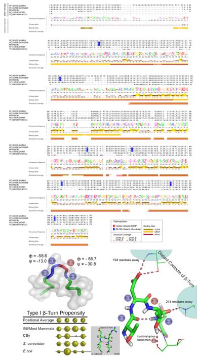 Fig. 1 Structural and sequence analysis of MSH3.1
Key structural properties of MSH3:
- Contains a classic ATP dual module combined with domain and mismatch recognition domain
- Form a clamp-like dimer interface to synergistically bind DNA
- Specific helix-turn-helix motifs are responsible for recognizing DNA insertion/deletion loops
Functions of MSH3
The core function of the MSH3 gene is to participate in DNA mismatch repair. However, this gene also plays a significant role in various cellular processes, including gene recombination regulation and cell cycle checkpoint responses.
| Function | Description |
| Insertion/missing repair | Specifically identify and correct 1-16 nucleotide insertion/deletion loops generated during DNA replication to maintain genomic stability. |
| Regulation of homologous recombination | In the fracture repair double-stranded DNA auxiliary homologous recombination process, to ensure accurate chromosome exchange. |
| Trinucleotide repeats are stable | Prevent abnormal amplification of trinucleotide repeat fragments such as Huntington's disease-related sequences. |
| Cell cycle regulation | By participating in the response to DNA damage checkpoints, it influences the cell cycle process and apoptotic decisions. |
| Association with chemotherapy resistance | There is a significant correlation between the expression level and the therapeutic sensitivity of some tumors to platinum drugs. |
The MutSβ complex formed by MSH3 and MSH2 demonstrates substrate specificity distinct from other mismatch repair proteins. Its functional loss mainly leads to insertion/deletion instability of microsatellite sequences rather than single nucleotide variations, which reflects its unique role in maintaining stability in specific genomic regions.
Applications of MSH3 and MSH3 Antibody in Literature
1. O'Reilly, Daniel, et al. "Di-valent siRNA-mediated silencing of MSH3 blocks somatic repeat expansion in mouse models of Huntington's disease." Molecular Therapy 31.6 (2023): 1661-1674. https://doi.org/10.1016/j.ymthe.2023.05.006
This study found that a chemically modified siRNA can effectively silence the MSH3 gene, thereby blocking the somatic expansion of CAG repeat sequences in the Huntington's disease mouse model without affecting other functions of the mismatch repair system, providing a new strategy for the treatment of this type of disease.
2. Oh, Jung-Min, et al. "MSH2-MSH3 promotes DNA end resection during homologous recombination and blocks polymerase theta-mediated end-joining through interaction with SMARCAD1 and EXO1." Nucleic acids research 51.11 (2023): 5584-5602. https://doi.org/10.1093/nar/gkad308
Research has found that the MSH2-MSH3 complex is recruited to the double-strand breaks of DNA through interaction with SMARCAD1. It guides repair towards homologous recombination rather than error-prone terminal connection pathways by promoting exo1-mediated long-range terminal excision and inhibiting the POLθ pathway.
3. Medina-Rivera, Melisa, et al. "Elevated MSH2 MSH3 expression interferes with DNA metabolism in vivo." Nucleic Acids Research 51.22 (2023): 12185-12206. https://doi.org/10.1093/nar/gkad934
Studies have shown that abnormally elevated levels of the MSH2-MSH3 complex can actively interfere with DNA replication and base excision repair through its ATPase activity, and cause cell cycle arrest. This reveals the crucial impact of MSH2-MSH3 protein abundance on its dual role in genomic stability.
4. Aelvoet, Arthur S., et al. "A large family with MSH3-related polyposis." Familial Cancer 22.1 (2023): 49-54. https://doi.org/10.1007/s10689-022-00297-x
Research has found that the biallelic pathogenic variation of the MSH3 gene is a rare cause of attenuated adenomatous polyposis. It was confirmed in a large family that this variation can cause colorectal adenomatous polyposis that occurs in middle age, and the EMAST phenotype was detected in both tumors and normal mucosa.
5. Koi, Minoru, et al. "Compound heterozygous MSH3 germline variants and associated tumor somatic DNA mismatch repair dysfunction." NPJ Precision Oncology 8.1 (2024): 12. https://doi.org/10.1038/s41698-024-00511-2
This study reports the fourth case caused by a biallelic germline variation of the MSH3 gene. The patient was diagnosed with colon cancer in middle age, and the tumor presented EMAST characteristics. Research has found that the mutated MSH3 protein is abnormally located on the cytoplasm and membrane, unable to enter the nucleus, and may induce tumorigenesis by interfering with the formation of the MSH2-MSH6 complex.
Creative Biolabs: MSH3 Antibodies for Research
Creative Biolabs specializes in the production of high-quality MSH3 antibodies for research and industrial applications. Our portfolio includes monoclonal antibodies tailored for ELISA, Flow Cytometry, Western blot, immunohistochemistry, and other diagnostic methodologies.
- Custom MSH3 Antibody Development: Tailor-made solutions to meet specific research requirements.
- Bulk Production: Large-scale antibody manufacturing for industry partners.
- Technical Support: Expert consultation for protocol optimization and troubleshooting.
- Aliquoting Services: Conveniently sized aliquots for long-term storage and consistent experimental outcomes.
For more details on our MSH3 antibodies, custom preparations, or technical support, contact us at email.
Reference
- Tomé, Stéphanie, et al. "MSH3 polymorphisms and protein levels affect CAG repeat instability in Huntington's disease mice." PLoS genetics 9.2 (2013): e1003280. https://doi.org/10.1371/journal.pgen.1003280
Anti-MSH3 antibodies
 Loading...


Hot products 
-
Mouse Anti-ATG5 Recombinant Antibody (9H197) (CBMAB-A3945-YC)

-
Rabbit Anti-ADRA1A Recombinant Antibody (V2-12532) (CBMAB-1022-CN)

-
Mouse Anti-ARID1B Recombinant Antibody (KMN1) (CBMAB-A3546-YC)

-
Rabbit Anti-ALK (Phosphorylated Y1278) Recombinant Antibody (D59G10) (PTM-CBMAB-0035YC)

-
Mouse Anti-BRCA2 Recombinant Antibody (CBYY-1728) (CBMAB-2077-YY)

-
Mouse Anti-BIRC5 Recombinant Antibody (6E4) (CBMAB-CP2646-LY)

-
Mouse Anti-CORO1A Recombinant Antibody (4G10) (V2LY-1206-LY806)

-
Mouse Anti-dsRNA Recombinant Antibody (2) (CBMAB-D1807-YC)

-
Mouse Anti-ADAM12 Recombinant Antibody (V2-179752) (CBMAB-A1114-YC)

-
Armenian hamster Anti-CD40 Recombinant Antibody (HM40-3) (CBMAB-C10365-LY)

-
Mouse Anti-ACTG1 Recombinant Antibody (V2-179597) (CBMAB-A0916-YC)

-
Mouse Anti-BSN Recombinant Antibody (219E1) (CBMAB-1228-CN)

-
Mouse Anti-CAPZB Recombinant Antibody (CBYY-C0944) (CBMAB-C2381-YY)

-
Mouse Anti-AAV9 Recombinant Antibody (V2-634029) (CBMAB-AP023LY)

-
Mouse Anti-CALR Recombinant Antibody (CBFYC-0763) (CBMAB-C0818-FY)

-
Mouse Anti-AFDN Recombinant Antibody (V2-58751) (CBMAB-L0408-YJ)

-
Rabbit Anti-CAMK2A Recombinant Antibody (BA0032) (CBMAB-0137CQ)

-
Mouse Anti-BANF1 Recombinant Antibody (3F10-4G12) (CBMAB-A0707-LY)

-
Mouse Anti-ADIPOR1 Recombinant Antibody (V2-179982) (CBMAB-A1368-YC)

-
Mouse Anti-AKR1C3 Recombinant Antibody (V2-12560) (CBMAB-1050-CN)

- AActivation
- AGAgonist
- APApoptosis
- BBlocking
- BABioassay
- BIBioimaging
- CImmunohistochemistry-Frozen Sections
- CIChromatin Immunoprecipitation
- CTCytotoxicity
- CSCostimulation
- DDepletion
- DBDot Blot
- EELISA
- ECELISA(Cap)
- EDELISA(Det)
- ESELISpot
- EMElectron Microscopy
- FFlow Cytometry
- FNFunction Assay
- GSGel Supershift
- IInhibition
- IAEnzyme Immunoassay
- ICImmunocytochemistry
- IDImmunodiffusion
- IEImmunoelectrophoresis
- IFImmunofluorescence
- IGImmunochromatography
- IHImmunohistochemistry
- IMImmunomicroscopy
- IOImmunoassay
- IPImmunoprecipitation
- ISIntracellular Staining for Flow Cytometry
- LALuminex Assay
- LFLateral Flow Immunoassay
- MMicroarray
- MCMass Cytometry/CyTOF
- MDMeDIP
- MSElectrophoretic Mobility Shift Assay
- NNeutralization
- PImmunohistologyp-Paraffin Sections
- PAPeptide Array
- PEPeptide ELISA
- PLProximity Ligation Assay
- RRadioimmunoassay
- SStimulation
- SESandwich ELISA
- SHIn situ hybridization
- TCTissue Culture
- WBWestern Blot
