PDPN Antibodies
Background
PDPN is a small molecule transmembrane glycoprotein that is mainly distributed on the surface of lymphatic endothelial cells and certain specific types of tumor cells. This protein participates in regulating lymphangiogenesis, platelet aggregation, and tumor metastasis processes, and plays a crucial role in lymph node metastasis of malignant tumors. It was first isolated and identified by Gunnarsson et al. from osteosarcoma cell lines in 2002. PDPN has attracted much attention due to its dual identity as a specific marker of lymphatic vessels and in tumor biology. Its structure is simple but its functions are diverse. Subsequent studies have continuously revealed its new roles in developmental biology, immune regulation, and thrombosis, providing important basis for tumor diagnosis, prognosis assessment, and targeted therapy.
Structure of PDPN
PDPN is a type I transmembrane glycoprotein with a molecular weight of approximately 36-40 kDa. There are variations in molecular weight among different species, mainly due to the degree of glycosylation and minor changes in the amino acid sequence.
The molecular weight of human PDPN is approximately 38 kDa and it is glycosylated; in mice, it is about 36 kDa, with slightly different glycosylation sites from those in humans; in rats, it is approximately 36-38 kDa, and the sequence is highly similar to that of humans; in dogs, it is about 37 kDa, with relatively conserved structure; in rabbits, it is about 38 kDa, and the antibody cross-reactivity is good.
The extracellular region of the PDPN protein is rich in serine and threonine residues, which is the main area for O-glycosylation. Its transmembrane region is highly conserved, while the intracellular region is relatively short but contains multiple phosphorylation sites, which can mediate intracellular signal transduction. The platelet receptor CLEC-2 recognizes the specific glycosylation epitopes of PDPN and activates downstream signaling pathways. This protein is stably expressed in lymphatic endothelial cells, but is abnormally highly expressed in some tumor cells, often indicating an increased risk of lymph node metastasis.
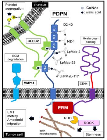 Fig. 1 Schematic representation of podoplanin (PDPN) structure and functions.1
Key structural properties of PDPN:
- Type I transmembrane glycoprotein structure, with a longer extracellular region
- The O-glycosylation region rich in serine/threonine
- Across the membrane area highly conservative, intracellular shorter tail area
- Nearly with CLEC - 2 receptor recognition with platelet membrane end table
Functions of PDPN
The main function of PDPN is to participate in lymphangiogenesis and tumor metastasis, but it also plays a role in various physiological and pathological processes, including platelet aggregation, immune regulation and developmental signal transduction.
| Function | Description |
| Lymphangiogenesis | PDPN is expressed on the surface of lymphatic endothelial cells and regulates the formation and remodeling of the lymphatic network. |
| Tumor metastasis | Highly expressed in various tumor cells, it promotes cancer cell migration and lymph node metastasis by interacting with the CLEC-2 receptor. |
| Platelet aggregation | The PDPN on the surface of tumor cells can induce platelet aggregation, forming microthrombi and assisting the tumor in surviving in the circulatory system. |
| Immune regulation | Inducibly expressed on immune cells such as macrophages, it participates in inflammatory responses and fibrosis processes. |
| Organ development | Temporarily expressed during the development of organs such as the lung, kidney, and heart, it participates in epithelial-mesenchymal interactions. |
The binding of PDPN to its endogenous ligand CLEC-2 is highly specific. This axis is involved in the maturation of cerebral blood vessels and lymphatic vessel separation during embryonic development, and is largely silent in adulthood. However, it is reactivated in various malignant tumors.
Applications of PDPN and PDPN Antibody in Literature
1. Suzuki, Hiroyuki, Mika K. Kaneko, and Yukinari Kato. "Roles of podoplanin in malignant progression of tumor." Cells 11.3 (2022): 575. https://doi.org/10.3390/cells11030575
Studies have shown that PDPN is a glycoprotein that is overexpressed in various tumors, which can promote tumor malignancy progression and immune evasion, and participate in the remodeling of the tumor microenvironment. Targeted therapeutic strategies against PDPN have become a new research direction in cancer treatment.
2. Sikorska, Justyna, et al. "Podoplanin (PDPN) affects the invasiveness of thyroid carcinoma cells by inducing ezrin, radixin and moesin (E/R/M) phosphorylation in association with matrix metalloproteinases." BMC cancer 19.1 (2019): 85. https://doi.org/10.1186/s12885-018-5239-z
The research shows that in thyroid cancer BcPAP cells, inhibiting the expression of PDPN can reduce cell migration and invasion by regulating E/R/M proteins and MMPs, and its effect is related to the MAPK signaling pathway.
3. Feng, Chunyan, et al. "A novel PDPN antagonist peptide CY12-RP2 inhibits melanoma growth via Wnt/β-catenin and modulates the immune cells." Journal of Experimental & Clinical Cancer Research 43.1 (2024): 9. https://doi.org/10.1186/s13046-023-02910-y
Studies have shown that PDPN is overexpressed in melanoma. Its antagonist peptide CY12-RP2 can specifically bind to PDPN and effectively inhibit tumor growth and metastasis by suppressing the Wnt/β-catenin pathway and activating immune cells.
4. Wang, Xuya, et al. "PDPN contributes to constructing immunosuppressive microenvironment in IDH wildtype glioma." Cancer Gene Therapy 30.2 (2023): 345-357. https://doi.org/10.1038/s41417-022-00550-6
Studies have confirmed that PDPN is highly expressed in IDH wild-type gliomas and is an independent poor prognostic indicator. It promotes the M2 polarization of macrophages and the degranulation of neutrophils, thereby driving the formation of an immunosuppressive microenvironment.
5. Li, Ke, et al. "A circular RNA activated by TGFβ promotes tumor metastasis through enhancing IGF2BP3-mediated PDPN mRNA stability." Nature Communications 14.1 (2023): 6876. https://doi.org/10.1038/s41467-023-42571-1
The study found that TGFβ-induced circITGB6 stabilizes PDPN mRNA, enhancing the epithelial-mesenchymal transition and metastasis ability of colorectal cancer. Its high expression is associated with poor prognosis in patients. Targeting circITGB6 is expected to inhibit tumor metastasis.
Creative Biolabs: PDPN Antibodies for Research
Creative Biolabs specializes in the production of high-quality PDPN antibodies for research and industrial applications. Our portfolio includes monoclonal and polyclonal antibodies tailored for ELISA, Flow Cytometry, Western blot, immunohistochemistry, and other diagnostic methodologies.
- Custom PDPN Antibody Development: Tailor-made solutions to meet specific research requirements.
- Bulk Production: Large-scale antibody manufacturing for industry partners.
- Technical Support: Expert consultation for protocol optimization and troubleshooting.
- Aliquoting Services: Conveniently sized aliquots for long-term storage and consistent experimental outcomes.
For more details on our PDPN antibodies, custom preparations, or technical support, contact us at email.
Reference
- Suzuki, Hiroyuki, Mika K. Kaneko, and Yukinari Kato. "Roles of podoplanin in malignant progression of tumor." Cells 11.3 (2022): 575. Distributed under Open Access license CC BY 4.0, without modification. https://doi.org/10.3390/cells11030575
Anti-PDPN antibodies
 Loading...


Hot products 
-
Mouse Anti-ALX1 Recombinant Antibody (96k) (CBMAB-C0616-FY)

-
Mouse Anti-HTLV-1 gp46 Recombinant Antibody (CBMW-H1006) (CBMAB-V208-1154-FY)

-
Rabbit Anti-AP2M1 (Phosphorylated T156) Recombinant Antibody (D4F3) (PTM-CBMAB-0610LY)

-
Rabbit Anti-AKT2 (Phosphorylated S474) Recombinant Antibody (V2-556130) (PTM-CBMAB-0605LY)

-
Rabbit Anti-CCN1 Recombinant Antibody (CBWJC-3580) (CBMAB-C4816WJ)

-
Mouse Anti-CD24 Recombinant Antibody (SN3) (CBMAB-C1037-CQ)

-
Mouse Anti-CARD11 Recombinant Antibody (CBFYC-0811) (CBMAB-C0866-FY)

-
Mouse Anti-BIRC5 Recombinant Antibody (6E4) (CBMAB-CP2646-LY)

-
Mouse Anti-ATM Recombinant Antibody (2C1) (CBMAB-A3970-YC)

-
Mouse Anti-APOE Recombinant Antibody (A1) (CBMAB-0078CQ)

-
Mouse Anti-BCL6 Recombinant Antibody (CBYY-0435) (CBMAB-0437-YY)

-
Mouse Anti-CASQ1 Recombinant Antibody (CBFYC-0863) (CBMAB-C0918-FY)

-
Mouse Anti-CCND2 Recombinant Antibody (DCS-3) (CBMAB-G1318-LY)

-
Mouse Anti-CDK7 Recombinant Antibody (CBYY-C1783) (CBMAB-C3221-YY)

-
Rabbit Anti-CBL Recombinant Antibody (D4E10) (CBMAB-CP0149-LY)

-
Rat Anti-AChR Recombinant Antibody (V2-12500) (CBMAB-0990-CN)

-
Mouse Anti-AAV8 Recombinant Antibody (V2-634028) (CBMAB-AP022LY)

-
Rabbit Anti-ABL1 (Phosphorylated Y245) Recombinant Antibody (V2-505716) (PTM-CBMAB-0465LY)

-
Mouse Anti-ALB Recombinant Antibody (V2-180650) (CBMAB-A2186-YC)

-
Mouse Anti-AAV-5 Recombinant Antibody (V2-503417) (CBMAB-V208-1369-FY)

- AActivation
- AGAgonist
- APApoptosis
- BBlocking
- BABioassay
- BIBioimaging
- CImmunohistochemistry-Frozen Sections
- CIChromatin Immunoprecipitation
- CTCytotoxicity
- CSCostimulation
- DDepletion
- DBDot Blot
- EELISA
- ECELISA(Cap)
- EDELISA(Det)
- ESELISpot
- EMElectron Microscopy
- FFlow Cytometry
- FNFunction Assay
- GSGel Supershift
- IInhibition
- IAEnzyme Immunoassay
- ICImmunocytochemistry
- IDImmunodiffusion
- IEImmunoelectrophoresis
- IFImmunofluorescence
- IGImmunochromatography
- IHImmunohistochemistry
- IMImmunomicroscopy
- IOImmunoassay
- IPImmunoprecipitation
- ISIntracellular Staining for Flow Cytometry
- LALuminex Assay
- LFLateral Flow Immunoassay
- MMicroarray
- MCMass Cytometry/CyTOF
- MDMeDIP
- MSElectrophoretic Mobility Shift Assay
- NNeutralization
- PImmunohistologyp-Paraffin Sections
- PAPeptide Array
- PEPeptide ELISA
- PLProximity Ligation Assay
- RRadioimmunoassay
- SStimulation
- SESandwich ELISA
- SHIn situ hybridization
- TCTissue Culture
- WBWestern Blot
